7ZV3

Crystal structure of indoleamine 2,3-dioxygenase 1 (IDO1) in complex with ferric heme and MMG-0472


Experimental Data Snapshot

  • Method: X-RAY DIFFRACTION
  • Resolution: 2.55 Å
  • R-Value Free: 0.241 
  • R-Value Work: 0.191 
  • R-Value Observed: 0.194 

wwPDB Validation   3D Report Full Report


Ligand Structure Quality Assessment 


This is version 1.2 of the entry. See complete history


Literature

Structure-based optimization of type III indoleamine 2,3-dioxygenase 1 (IDO1) inhibitors.

Rohrig, U.F.Majjigapu, S.R.Vogel, P.Reynaud, A.Pojer, F.Dilek, N.Reichenbach, P.Ascencao, K.Irving, M.Coukos, G.Michielin, O.Zoete, V.

(2022) J Enzyme Inhib Med Chem 37: 1773-1811

  • DOI: https://doi.org/10.1080/14756366.2022.2089665
  • Primary Citation of Related Structures:  
    7ZV3

  • PubMed Abstract: 

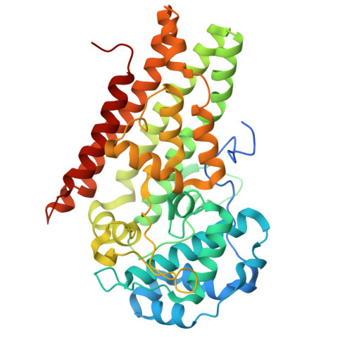
    The haem enzyme indoleamine 2,3-dioxygenase 1 (IDO1) catalyses the rate-limiting step in the kynurenine pathway of tryptophan metabolism and plays an essential role in immunity, neuronal function, and ageing. Expression of IDO1 in cancer cells results in the suppression of an immune response, and therefore IDO1 inhibitors have been developed for use in anti-cancer immunotherapy. Here, we report an extension of our previously described highly efficient haem-binding 1,2,3-triazole and 1,2,4-triazole inhibitor series, the best compound having both enzymatic and cellular IC 50 values of 34 nM. We provide enzymatic inhibition data for almost 100 new compounds and X-ray diffraction data for one compound in complex with IDO1. Structural and computational studies explain the dramatic drop in activity upon extension to pocket B, which has been observed in diverse haem-binding inhibitor scaffolds. Our data provides important insights for future IDO1 inhibitor design.


  • Organizational Affiliation

    SIB Swiss Institute of Bioinformatics, Molecular Modeling Group, Lausanne, Switzerland.


Macromolecules
Find similar proteins by:  (by identity cutoff)  |  3D Structure
Entity ID: 1
MoleculeChains Sequence LengthOrganismDetailsImage
Indoleamine 2,3-dioxygenase 1
A, B
423Homo sapiensMutation(s): 0 
Gene Names: IDO1IDOINDO
EC: 1.13.11.52
UniProt & NIH Common Fund Data Resources
Find proteins for P14902 (Homo sapiens)
Explore P14902 
Go to UniProtKB:  P14902
PHAROS:  P14902
GTEx:  ENSG00000131203 
Entity Groups  
Sequence Clusters30% Identity50% Identity70% Identity90% Identity95% Identity100% Identity
UniProt GroupP14902
Sequence Annotations
Expand
  • Reference Sequence
Small Molecules
Ligands 4 Unique
IDChains Name / Formula / InChI Key2D Diagram3D Interactions
HEM
Query on HEM

Download Ideal Coordinates CCD File 
C [auth A],
G [auth B]
PROTOPORPHYRIN IX CONTAINING FE
C34 H32 Fe N4 O4
KABFMIBPWCXCRK-RGGAHWMASA-L
RCT (Subject of Investigation/LOI)
Query on RCT

Download Ideal Coordinates CCD File 
D [auth A],
H [auth B]
4-(3-chloro-2-phenethylphenyl)-1H-1,2,3-triazole
C16 H14 Cl N3
KSCUUNCHIFGAIO-UHFFFAOYSA-N
SO4
Query on SO4

Download Ideal Coordinates CCD File 
F [auth A],
I [auth B]
SULFATE ION
O4 S
QAOWNCQODCNURD-UHFFFAOYSA-L
GOL
Query on GOL

Download Ideal Coordinates CCD File 
E [auth A]GLYCEROL
C3 H8 O3
PEDCQBHIVMGVHV-UHFFFAOYSA-N
Experimental Data & Validation

Experimental Data

  • Method: X-RAY DIFFRACTION
  • Resolution: 2.55 Å
  • R-Value Free: 0.241 
  • R-Value Work: 0.191 
  • R-Value Observed: 0.194 
  • Space Group: P 21 21 21
Unit Cell:
Length ( Å )Angle ( ˚ )
a = 86.65α = 90
b = 97.419β = 90
c = 131.996γ = 90
Software Package:
Software NamePurpose
PHENIXrefinement
XDSdata reduction
XDSdata scaling
PHASERphasing

Structure Validation

View Full Validation Report


Ligand Structure Quality Assessment 


Entry History & Funding Information

Deposition Data


Funding OrganizationLocationGrant Number
Not funded--

Revision History  (Full details and data files)

  • Version 1.0: 2022-07-06
    Type: Initial release
  • Version 1.1: 2024-01-31
    Changes: Data collection, Refinement description
  • Version 1.2: 2024-03-27
    Changes: Database references