7ZCX

S-layer protein SlaA from Sulfolobus acidocaldarius at pH 4.0


Experimental Data Snapshot

  • Method: ELECTRON MICROSCOPY
  • Resolution: 3.10 Å
  • Aggregation State: PARTICLE 
  • Reconstruction Method: SINGLE PARTICLE 

wwPDB Validation   3D Report Full Report


This is version 2.0 of the entry. See complete history


Literature

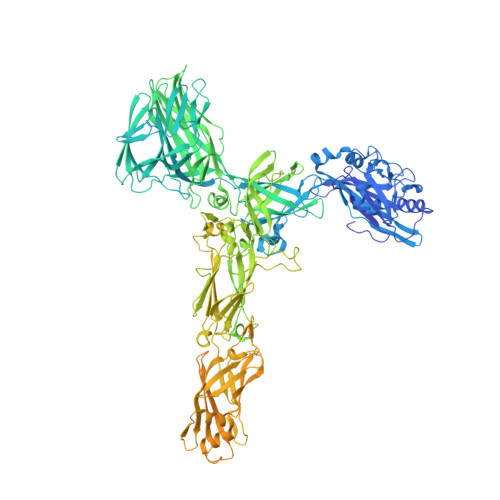
Structure of the two-component S-layer of the archaeon Sulfolobus acidocaldarius.

Gambelli, L.McLaren, M.Conners, R.Sanders, K.Gaines, M.C.Clark, L.Gold, V.A.M.Kattnig, D.Sikora, M.Hanus, C.Isupov, M.N.Daum, B.

(2024) Elife 13

  • DOI: https://doi.org/10.7554/eLife.84617
  • Primary Citation of Related Structures:  
    7ZCX, 8AN2, 8AN3, 8QOX, 8QP0

  • PubMed Abstract: 

    Surface layers (S-layers) are resilient two-dimensional protein lattices that encapsulate many bacteria and most archaea. In archaea, S-layers usually form the only structural component of the cell wall and thus act as the final frontier between the cell and its environment. Therefore, S-layers are crucial for supporting microbial life. Notwithstanding their importance, little is known about archaeal S-layers at the atomic level. Here, we combined single-particle cryo electron microscopy, cryo electron tomography, and Alphafold2 predictions to generate an atomic model of the two-component S-layer of Sulfolobus acidocaldarius . The outer component of this S-layer (SlaA) is a flexible, highly glycosylated, and stable protein. Together with the inner and membrane-bound component (SlaB), they assemble into a porous and interwoven lattice. We hypothesise that jackknife-like conformational changes in SlaA play important roles in S-layer assembly.


  • Organizational Affiliation

    Living Systems Institute, University of Exeter, Exeter, United Kingdom.


Macromolecules
Find similar proteins by:  (by identity cutoff)  |  3D Structure
Entity ID: 1
MoleculeChains Sequence LengthOrganismDetailsImage
S-layer protein AA [auth AAA]1,424Sulfolobus acidocaldariusMutation(s): 0 
Gene Names: slaAslp1Saci_2355
UniProt
Find proteins for Q4J6E5 (Sulfolobus acidocaldarius (strain ATCC 33909 / DSM 639 / JCM 8929 / NBRC 15157 / NCIMB 11770))
Explore Q4J6E5 
Go to UniProtKB:  Q4J6E5
Entity Groups  
Sequence Clusters30% Identity50% Identity70% Identity90% Identity95% Identity100% Identity
UniProt GroupQ4J6E5
Sequence Annotations
Expand
  • Reference Sequence
Oligosaccharides

Help

Entity ID: 2
MoleculeChains Length2D Diagram Glycosylation3D Interactions
6-deoxy-6-sulfo-beta-D-glucopyranose-(1-3)-[alpha-D-mannopyranose-(1-6)]2-acetamido-2-deoxy-beta-D-glucopyranose-(1-4)-2-acetamido-2-deoxy-beta-D-glucopyranoseB [auth aaa],
F [auth D],
P [auth N]
4N/AN-Glycosylation
Entity ID: 3
MoleculeChains Length2D Diagram Glycosylation3D Interactions
2-acetamido-2-deoxy-beta-D-glucopyranose-(1-4)-2-acetamido-2-deoxy-beta-D-glucopyranoseC [auth A],
L [auth J]
2N-Glycosylation
Glycosylation Resources
GlyTouCan:  G42666HT
GlyCosmos:  G42666HT
GlyGen:  G42666HT
Entity ID: 4
MoleculeChains Length2D Diagram Glycosylation3D Interactions
beta-D-glucopyranose-(1-4)-6-deoxy-6-sulfo-beta-D-glucopyranose-(1-3)-[alpha-D-mannopyranose-(1-4)][alpha-D-mannopyranose-(1-6)]2-acetamido-2-deoxy-beta-D-glucopyranose-(1-4)-2-acetamido-2-deoxy-beta-D-glucopyranoseD [auth B],
H [auth F],
N [auth L]
6N/AN-Glycosylation
Entity ID: 5
MoleculeChains Length2D Diagram Glycosylation3D Interactions
6-deoxy-6-sulfo-beta-D-glucopyranose-(1-3)-[alpha-D-mannopyranose-(1-4)][alpha-D-mannopyranose-(1-6)]2-acetamido-2-deoxy-beta-D-glucopyranose-(1-4)-2-acetamido-2-deoxy-beta-D-glucopyranose
E [auth C],
G [auth E],
I [auth G],
J [auth H],
K [auth I],
E [auth C],
G [auth E],
I [auth G],
J [auth H],
K [auth I],
O [auth M],
Q [auth O],
S [auth Q]
5N/AN-Glycosylation
Entity ID: 6
MoleculeChains Length2D Diagram Glycosylation3D Interactions
alpha-D-mannopyranose-(1-6)-2-acetamido-2-deoxy-beta-D-glucopyranose-(1-4)-2-acetamido-2-deoxy-beta-D-glucopyranoseM [auth K]3N-Glycosylation
Glycosylation Resources
GlyTouCan:  G34031ES
GlyCosmos:  G34031ES
GlyGen:  G34031ES
Entity ID: 7
MoleculeChains Length2D Diagram Glycosylation3D Interactions
alpha-D-mannopyranose-(1-4)-[alpha-D-mannopyranose-(1-6)]2-acetamido-2-deoxy-beta-D-glucopyranose-(1-4)-2-acetamido-2-deoxy-beta-D-glucopyranoseR [auth P]4N-Glycosylation
Small Molecules
Ligands 2 Unique
IDChains Name / Formula / InChI Key2D Diagram3D Interactions
YZT (Subject of Investigation/LOI)
Query on YZT

Download Ideal Coordinates CCD File 
U [auth AAA]6-deoxy-6-sulfo-beta-D-glucopyranose
C6 H12 O8 S
QFBWOLBPVQLZEH-VFUOTHLCSA-N
NAG (Subject of Investigation/LOI)
Query on NAG

Download Ideal Coordinates CCD File 
T [auth AAA]2-acetamido-2-deoxy-beta-D-glucopyranose
C8 H15 N O6
OVRNDRQMDRJTHS-FMDGEEDCSA-N
Experimental Data & Validation

Experimental Data

  • Method: ELECTRON MICROSCOPY
  • Resolution: 3.10 Å
  • Aggregation State: PARTICLE 
  • Reconstruction Method: SINGLE PARTICLE 
EM Software:
TaskSoftware PackageVersion
MODEL REFINEMENTREFMAC

Structure Validation

View Full Validation Report


Entry History & Funding Information

Deposition Data


Funding OrganizationLocationGrant Number
European Research Council (ERC)European Union803894

Revision History  (Full details and data files)

  • Version 1.0: 2023-06-14
    Type: Initial release
  • Version 2.0: 2024-02-21
    Changes: Atomic model, Data collection, Database references, Derived calculations, Refinement description