7Z32

Escherichia coli periplasmic phytase AppA D304A mutant, phosphohistidine intermediate


Experimental Data Snapshot

  • Method: X-RAY DIFFRACTION
  • Resolution: 1.85 Å
  • R-Value Free: 0.224 
  • R-Value Work: 0.163 
  • R-Value Observed: 0.166 

wwPDB Validation   3D Report Full Report


This is version 1.2 of the entry. See complete history


Literature

Insights to the Structural Basis for the Stereospecificity of the Escherichia coli Phytase, AppA.

Acquistapace, I.M.Thompson, E.J.Kuhn, I.Bedford, M.R.Brearley, C.A.Hemmings, A.M.

(2022) Int J Mol Sci 23

  • DOI: https://doi.org/10.3390/ijms23116346
  • Primary Citation of Related Structures:  
    7Z1J, 7Z2S, 7Z2T, 7Z2W, 7Z2Y, 7Z32, 7Z3V

  • PubMed Abstract: 

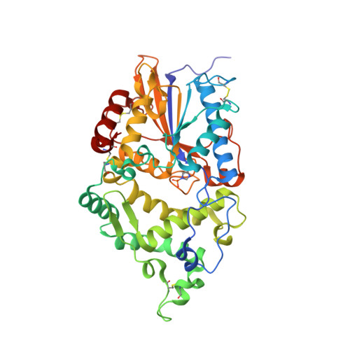
    AppA, the Escherichia coli periplasmic phytase of clade 2 of the histidine phosphatase (HP2) family, has been well-characterized and successfully engineered for use as an animal feed supplement. AppA is a 1D-6-phytase and highly stereospecific but transiently accumulates 1D- myo -Ins(2,3,4,5)P 4 and other lower phosphorylated intermediates. If this bottleneck in liberation of orthophosphate is to be obviated through protein engineering, an explanation of its rather rigid preference for the initial site and subsequent cleavage of phytic acid is required. To help explain this behaviour, the role of the catalytic proton donor residue in determining AppA stereospecificity was investigated. Four variants were generated by site-directed mutagenesis of the active site HDT amino acid sequence motif containing the catalytic proton donor, D304. The identity and position of the prospective proton donor residue was found to strongly influence stereospecificity. While the wild-type enzyme has a strong preference for 1D-6-phytase activity, a marked reduction in stereospecificity was observed for a D304E variant, while a proton donor-less mutant (D304A) displayed exclusive 1D-1/3-phytase activity. High-resolution X-ray crystal structures of complexes of the mutants with a non-hydrolysable substrate analogue inhibitor point to a crucial role played by D304 in stereospecificity by influencing the size and polarity of specificity pockets A and B. Taken together, these results provide the first evidence for the involvement of the proton donor residue in determining the stereospecificity of HP2 phytases and prepares the ground for structure-informed engineering studies targeting the production of animal feed enzymes capable of the efficient and complete dephosphorylation of dietary phytic acid.


  • Organizational Affiliation

    School of Biological Sciences, University of East Anglia, Norwich Research Park, Norwich NR4 7TJ, UK.


Macromolecules
Find similar proteins by:  (by identity cutoff)  |  3D Structure
Entity ID: 1
MoleculeChains Sequence LengthOrganismDetailsImage
Acidphosphatase412Escherichia coliMutation(s): 1 
Gene Names: 
appAappA_1appA_2A8C65_14190ABE90_015325ACN81_22175ACU57_13335AM464_22895AML23_15750AT845_000200B7C15_002169BANRA_01347BANRA_01497BANRA_01938BANRA_04663BHS87_05220BJI68_21075BJJ90_16615BO068_002094BON73_13465BON93_08495BON98_05845BTQ06_12390BUE81_23535BvCms2454_01079BvCmsHHP019_04940BvCmsHHP056_02151BvCmsKKP061_02619BvCmsKSNP073_03019BvCmsKSP011_04200BvCmsKSP067_04712C2U48_04610C3F40_07155C5F72_16390C5N07_03615C5Y87_10555C6N50_004791C7B02_01415C9160_17160CA593_23620CG831_002343D0X26_04185D3O91_18420D3Y67_02885D5H22_11490D9D77_12200D9J11_05545DAH34_01475DAH37_00935DEN89_03775DEN95_10355DIV22_11465DM968_16120DRW19_20065DTL43_09820DXT71_05565DXT73_09385E2119_16940E2135_21350E4K51_09405E4K54_00245EAI46_03420EC1094V2_2850EC95NR1_05304EGC08_19360EI041_12345EIZ93_00240ERS139208_02859ETECE36_03929ETECE925_02991F2N31_24545F9400_05360F9407_14150F9V24_04715F9X20_013415F9X20_13515FDM60_03565FOI11_008030FOI11_12015FQE77_13130FQF29_10640FV293_21365G4A38_18295G5632_05905G7635_003041GBE29_03585GF646_03565GKF89_10795GKG12_05120GP662_14105GQE64_05300GQE87_11110GQM04_09625GQM06_15635GRQ19_09290GRW05_05690GRW57_05130GRW80_07395GRW81_11950H4P50_16180H4P51_16030HJN04_004452HJO75_001864HMS79_12845HMV95_15325HNC36_01680HNC52_01435HPE39_08845HVV53_00565HVY77_16815HVZ33_15560HX136_16165HZ71_004423I6H02_23730NCTC11022_00303NCTC11181_00321NCTC13216_03584NCTC8008_02712NCTC8960_00665NCTC9048_03264NCTC9706_00521NCTC9962_02002SAMEA3472044_00046SAMEA3472064_00229SAMEA3472080_00519SAMEA3484427_04507SAMEA3484429_04453SAMEA3751407_00914SAMEA3752386_03823SAMEA3752557_01451SAMEA3753300_02779WP2S18E08_29460WQ89_13085WR15_11565

EC: 3.1.3.26 (PDB Primary Data), 3.1.3.2 (PDB Primary Data)
UniProt
Find proteins for P07102 (Escherichia coli (strain K12))
Explore P07102 
Go to UniProtKB:  P07102
Entity Groups  
Sequence Clusters30% Identity50% Identity70% Identity90% Identity95% Identity100% Identity
UniProt GroupP07102
Sequence Annotations
Expand
  • Reference Sequence
Small Molecules
Ligands 1 Unique
IDChains Name / Formula / InChI Key2D Diagram3D Interactions
NI
Query on NI

Download Ideal Coordinates CCD File 
B [auth A]NICKEL (II) ION
Ni
VEQPNABPJHWNSG-UHFFFAOYSA-N
Modified Residues  1 Unique
IDChains TypeFormula2D DiagramParent
NEP
Query on NEP
A
L-PEPTIDE LINKINGC6 H10 N3 O5 PHIS
Experimental Data & Validation

Experimental Data

  • Method: X-RAY DIFFRACTION
  • Resolution: 1.85 Å
  • R-Value Free: 0.224 
  • R-Value Work: 0.163 
  • R-Value Observed: 0.166 
  • Space Group: P 1 21 1
Unit Cell:
Length ( Å )Angle ( ˚ )
a = 63.75α = 90
b = 47.96β = 101.08
c = 65.97γ = 90
Software Package:
Software NamePurpose
PHENIXrefinement
PHENIXrefinement
xia2data reduction
pointlessdata scaling
PHASERphasing

Structure Validation

View Full Validation Report


Entry History & Funding Information

Deposition Data


Funding OrganizationLocationGrant Number
Biotechnology and Biological Sciences Research Council (BBSRC)United KingdomBB/M022978/1

Revision History  (Full details and data files)

  • Version 1.0: 2022-03-16
    Type: Initial release
  • Version 1.1: 2022-06-29
    Changes: Database references
  • Version 1.2: 2024-01-31
    Changes: Data collection, Refinement description