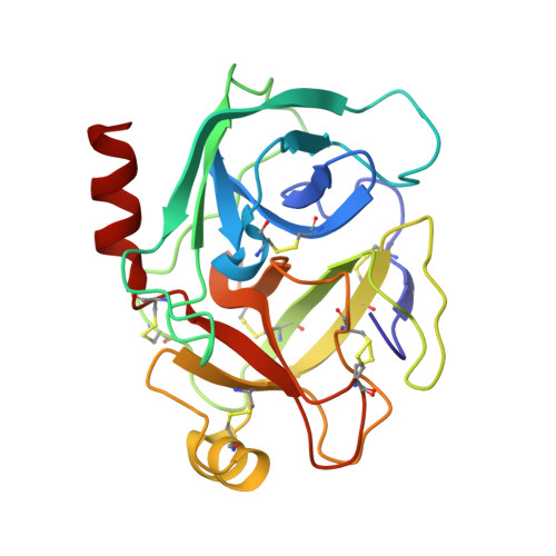
7Z2I

TRYPSIN (BOVINE) COMPLEXED WITH compound 4


Experimental Data Snapshot

  • Method: X-RAY DIFFRACTION
  • Resolution: 1.09 Å
  • R-Value Free: 0.150 
  • R-Value Work: 0.135 
  • R-Value Observed: 0.136 

wwPDB Validation   3D Report Full Report


Ligand Structure Quality Assessment 


This is version 1.3 of the entry. See complete history


Literature

Efficient Screening of Target-Specific Selected Compounds in Mixtures by 19 F NMR Binding Assay with Predicted 19 F NMR Chemical Shifts.

Vulpetti, A.Lingel, A.Dalvit, C.Schiering, N.Oberer, L.Henry, C.Lu, Y.

(2022) ChemMedChem 17: e202200163-e202200163

  • DOI: https://doi.org/10.1002/cmdc.202200163
  • Primary Citation of Related Structures:  
    7Z25, 7Z2I

  • PubMed Abstract: 

    Ligand-based 19 F NMR screening is a highly effective and well-established hit-finding approach. The high sensitivity to protein binding makes it particularly suitable for fragment screening. Different criteria can be considered for generating fluorinated fragment libraries. One common strategy is to assemble a large, diverse, well-designed and characterized fragment library which is screened in mixtures, generated based on experimental 19 F NMR chemical shifts. Here, we introduce a complementary knowledge-based 19 F NMR screening approach, named 19 Focused screening, enabling the efficient screening of putative active molecules selected by computational hit finding methodologies, in mixtures assembled and on-the-fly deconvoluted based on predicted 19 F NMR chemical shifts. In this study, we developed a novel approach, named LEFshift, for 19 F NMR chemical shift prediction using rooted topological fluorine torsion fingerprints in combination with a random forest machine learning method. A demonstration of this approach to a real test case is reported.


  • Organizational Affiliation

    Novartis Institutes for BioMedical Research, 4002, Basel, Switzerland.


Macromolecules
Find similar proteins by:  (by identity cutoff)  |  3D Structure
Entity ID: 1
MoleculeChains Sequence LengthOrganismDetailsImage
Serine protease 1223Bos taurusMutation(s): 0 
EC: 3.4.21.4
UniProt
Find proteins for P00760 (Bos taurus)
Explore P00760 
Go to UniProtKB:  P00760
Entity Groups  
Sequence Clusters30% Identity50% Identity70% Identity90% Identity95% Identity100% Identity
UniProt GroupP00760
Sequence Annotations
Expand
  • Reference Sequence
Experimental Data & Validation

Experimental Data

  • Method: X-RAY DIFFRACTION
  • Resolution: 1.09 Å
  • R-Value Free: 0.150 
  • R-Value Work: 0.135 
  • R-Value Observed: 0.136 
  • Space Group: P 21 21 21
Unit Cell:
Length ( Å )Angle ( ˚ )
a = 54.552α = 90
b = 58.299β = 90
c = 66.855γ = 90
Software Package:
Software NamePurpose
XSCALEdata scaling
REFMACrefinement
PDB_EXTRACTdata extraction
XDSdata reduction
MOLREPphasing

Structure Validation

View Full Validation Report


Ligand Structure Quality Assessment 


Entry History & Funding Information

Deposition Data


Funding OrganizationLocationGrant Number
Not funded--

Revision History  (Full details and data files)

  • Version 1.0: 2022-05-25
    Type: Initial release
  • Version 1.1: 2022-07-13
    Changes: Database references
  • Version 1.2: 2022-07-20
    Changes: Database references
  • Version 1.3: 2024-05-01
    Changes: Data collection, Refinement description