7YYW

Molecular snapshots of drug release from tubulin: 10 nanoseconds after photoactivation.


Experimental Data Snapshot

  • Method: X-RAY DIFFRACTION
  • Resolution: 2.20 Å
  • R-Value Free: 0.343 
  • R-Value Work: 0.311 
  • R-Value Observed: 0.312 

wwPDB Validation   3D Report Full Report


Ligand Structure Quality Assessment 


This is version 3.0 of the entry. See complete history


Literature

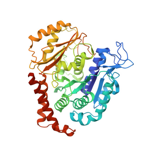
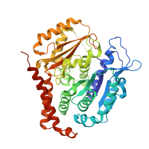
Watching the release of a photopharmacological drug from tubulin using time-resolved serial crystallography.

Wranik, M.Weinert, T.Slavov, C.Masini, T.Furrer, A.Gaillard, N.Gioia, D.Ferrarotti, M.James, D.Glover, H.Carrillo, M.Kekilli, D.Stipp, R.Skopintsev, P.Brunle, S.Muhlethaler, T.Beale, J.Gashi, D.Nass, K.Ozerov, D.Johnson, P.J.M.Cirelli, C.Bacellar, C.Braun, M.Wang, M.Dworkowski, F.Milne, C.Cavalli, A.Wachtveitl, J.Steinmetz, M.O.Standfuss, J.

(2023) Nat Commun 14: 903-903

  • DOI: https://doi.org/10.1038/s41467-023-36481-5
  • Primary Citation of Related Structures:  
    7YYQ, 7YYV, 7YYW, 7YYX, 7YYY, 7YYZ, 7YZ0, 7YZ1, 7YZ2, 7YZ3, 7YZ5, 7YZ6

  • PubMed Abstract: 

    The binding and release of ligands from their protein targets is central to fundamental biological processes as well as to drug discovery. Photopharmacology introduces chemical triggers that allow the changing of ligand affinities and thus biological activity by light. Insight into the molecular mechanisms of photopharmacology is largely missing because the relevant transitions during the light-triggered reaction cannot be resolved by conventional structural biology. Using time-resolved serial crystallography at a synchrotron and X-ray free-electron laser, we capture the release of the anti-cancer compound azo-combretastatin A4 and the resulting conformational changes in tubulin. Nine structural snapshots from 1 ns to 100 ms complemented by simulations show how cis-to-trans isomerization of the azobenzene bond leads to a switch in ligand affinity, opening of an exit channel, and collapse of the binding pocket upon ligand release. The resulting global backbone rearrangements are related to the action mechanism of microtubule-destabilizing drugs.


  • Organizational Affiliation

    Division of Biology and Chemistry, Paul Scherrer Institut, 5232, Villigen, Switzerland.


Macromolecules
Find similar proteins by:  (by identity cutoff)  |  3D Structure
Entity ID: 1
MoleculeChains Sequence LengthOrganismDetailsImage
Tubulin alpha-1B chain451Bos taurusMutation(s): 0 
UniProt
Find proteins for P81947 (Bos taurus)
Explore P81947 
Go to UniProtKB:  P81947
Entity Groups  
Sequence Clusters30% Identity50% Identity70% Identity90% Identity95% Identity100% Identity
UniProt GroupP81947
Sequence Annotations
Expand
  • Reference Sequence
Find similar proteins by:  (by identity cutoff)  |  3D Structure
Entity ID: 2
MoleculeChains Sequence LengthOrganismDetailsImage
Tubulin beta-2B chain445Bos taurusMutation(s): 0 
UniProt
Find proteins for Q6B856 (Bos taurus)
Explore Q6B856 
Go to UniProtKB:  Q6B856
Entity Groups  
Sequence Clusters30% Identity50% Identity70% Identity90% Identity95% Identity100% Identity
UniProt GroupQ6B856
Sequence Annotations
Expand
  • Reference Sequence
Find similar proteins by:  (by identity cutoff)  |  3D Structure
Entity ID: 3
MoleculeChains Sequence LengthOrganismDetailsImage
Designed Ankyrin Repeat Protein (DARPIN) D1C [auth F]169synthetic constructMutation(s): 0 
Entity Groups  
Sequence Clusters30% Identity50% Identity70% Identity90% Identity95% Identity100% Identity
Sequence Annotations
Expand
  • Reference Sequence
Small Molecules
Ligands 5 Unique
IDChains Name / Formula / InChI Key2D Diagram3D Interactions
GTP
Query on GTP

Download Ideal Coordinates CCD File 
D [auth A]GUANOSINE-5'-TRIPHOSPHATE
C10 H16 N5 O14 P3
XKMLYUALXHKNFT-UUOKFMHZSA-N
GDP
Query on GDP

Download Ideal Coordinates CCD File 
G [auth B]GUANOSINE-5'-DIPHOSPHATE
C10 H15 N5 O11 P2
QGWNDRXFNXRZMB-UUOKFMHZSA-N
VYT
Query on VYT

Download Ideal Coordinates CCD File 
H [auth B]Azo-Combretastatin A4 (trans)
C16 H18 N2 O5
AIRZFEKQGRSBNX-ISLYRVAYSA-N
CA
Query on CA

Download Ideal Coordinates CCD File 
F [auth A]CALCIUM ION
Ca
BHPQYMZQTOCNFJ-UHFFFAOYSA-N
MG
Query on MG

Download Ideal Coordinates CCD File 
E [auth A]MAGNESIUM ION
Mg
JLVVSXFLKOJNIY-UHFFFAOYSA-N
Experimental Data & Validation

Experimental Data

Unit Cell:
Length ( Å )Angle ( ˚ )
a = 74.53α = 90
b = 92.58β = 96.71
c = 83.99γ = 90
Software Package:
Software NamePurpose
PHENIXrefinement
PDB_EXTRACTdata extraction
CrystFELdata reduction
CrystFELdata scaling
PHASERphasing

Structure Validation

View Full Validation Report


Ligand Structure Quality Assessment 


Entry History & Funding Information

Deposition Data


Funding OrganizationLocationGrant Number
Swiss National Science FoundationSwitzerland31003A_179351
Swiss National Science FoundationSwitzerland310030_197674
Swiss National Science FoundationSwitzerland310030_192566
Swiss Nanoscience InstituteSwitzerlandSNI #1904
Other governmentNCCR:MUST
Other governmentProject WA 1850/4-3

Revision History  (Full details and data files)

  • Version 1.0: 2023-02-22
    Type: Initial release
  • Version 1.1: 2023-03-08
    Changes: Database references
  • Version 2.0: 2023-09-13
    Changes: Data collection, Non-polymer description, Structure summary
  • Version 3.0: 2023-09-27
    Changes: Atomic model, Data collection, Derived calculations, Non-polymer description, Structure summary