7YSR

GTPgammaS MT decorated with kinesin


Experimental Data Snapshot

  • Method: ELECTRON MICROSCOPY
  • Resolution: 4.30 Å
  • Aggregation State: HELICAL ARRAY 
  • Reconstruction Method: HELICAL 

wwPDB Validation   3D Report Full Report


This is version 1.0 of the entry. See complete history


Literature

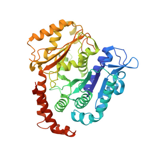
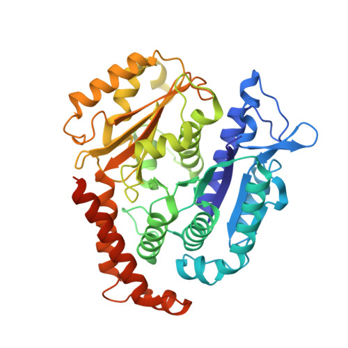
Structural insights into the mechanism of GTP initiation of microtubule assembly.

Zhou, J.Wang, A.Song, Y.Liu, N.Wang, J.Li, Y.Liang, X.Li, G.Chu, H.Wang, H.W.

(2023) Nat Commun 14: 5980-5980

  • DOI: https://doi.org/10.1038/s41467-023-41615-w
  • Primary Citation of Related Structures:  
    7YSN, 7YSO, 7YSP, 7YSQ, 7YSR

  • PubMed Abstract: 

    In eukaryotes, the dynamic assembly of microtubules (MT) plays an important role in numerous cellular processes. The underlying mechanism of GTP triggering MT assembly is still unknown. Here, we present cryo-EM structures of tubulin heterodimer at their GTP- and GDP-bound states, intermediate assembly states of GTP-tubulin, and final assembly stages of MT. Both GTP- and GDP-tubulin heterodimers adopt similar curved conformations with subtle flexibility differences. In head-to-tail oligomers of tubulin heterodimers, the inter-dimer interface of GDP-tubulin exhibits greater flexibility, particularly in tangential bending. Cryo-EM of the intermediate assembly states reveals two types of tubulin lateral contacts, "Tube-bond" and "MT-bond". Further, molecular dynamics (MD) simulations show that GTP triggers lateral contact formation in MT assembly in multiple sequential steps, gradually straightening the curved tubulin heterodimers. Therefore, we propose a flexible model of GTP-initiated MT assembly, including the formation of longitudinal and lateral contacts, to explain the nucleation and assembly of MT.


  • Organizational Affiliation

    State Key Laboratory of Membrane Biology, Tsinghua University, Beijing, 100084, China.


Macromolecules
Find similar proteins by:  (by identity cutoff)  |  3D Structure
Entity ID: 1
MoleculeChains Sequence LengthOrganismDetailsImage
Tubulin alpha chain
A, C
450Drosophila melanogasterMutation(s): 0 
UniProt
Find proteins for P06603 (Drosophila melanogaster)
Explore P06603 
Go to UniProtKB:  P06603
Entity Groups  
Sequence Clusters30% Identity50% Identity70% Identity90% Identity95% Identity100% Identity
UniProt GroupP06603
Sequence Annotations
Expand
  • Reference Sequence
Find similar proteins by:  (by identity cutoff)  |  3D Structure
Entity ID: 2
MoleculeChains Sequence LengthOrganismDetailsImage
Tubulin beta-1 chain
B, D
447Drosophila melanogasterMutation(s): 0 
UniProt
Find proteins for Q24560 (Drosophila melanogaster)
Explore Q24560 
Go to UniProtKB:  Q24560
Entity Groups  
Sequence Clusters30% Identity50% Identity70% Identity90% Identity95% Identity100% Identity
UniProt GroupQ24560
Sequence Annotations
Expand
  • Reference Sequence
Experimental Data & Validation

Experimental Data

  • Method: ELECTRON MICROSCOPY
  • Resolution: 4.30 Å
  • Aggregation State: HELICAL ARRAY 
  • Reconstruction Method: HELICAL 

Structure Validation

View Full Validation Report


Entry History & Funding Information

Deposition Data


Funding OrganizationLocationGrant Number
National Natural Science Foundation of China (NSFC)China31825009

Revision History  (Full details and data files)

  • Version 1.0: 2023-10-18
    Type: Initial release