


Crystal Structure of UDP-glucose 4-epimerase (Rv3634c) in complex with UDP-glucose in chainA and UDP-galactose in chain B from Mycobacterium tuberculosis
Yadav, S., Bhatia, I., Biswal, B.K.To be published.
Experimental Data Snapshot
Starting Model: experimental
View more details
Entity ID: 1 | |||||
|---|---|---|---|---|---|
| Molecule | Chains | Sequence Length | Organism | Details | Image |
| UDP-glucose 4-epimerase | 320 | Mycobacterium tuberculosis H37Rv | Mutation(s): 0 Gene Names: galE1 EC: 5.1.3.2 | 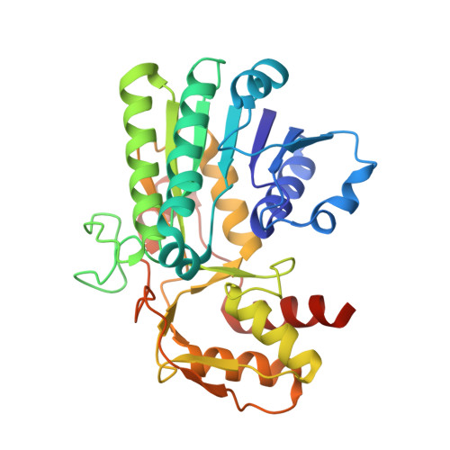 | |
UniProt | |||||
Entity Groups | |||||
| Sequence Clusters | 30% Identity50% Identity70% Identity90% Identity95% Identity100% Identity | ||||
| UniProt Group | P9WN67 | ||||
Sequence AnnotationsExpand | |||||
Reference Sequence | |||||
| Ligands 5 Unique | |||||
|---|---|---|---|---|---|
| ID | Chains | Name / Formula / InChI Key | 2D Diagram | 3D Interactions | |
| NAD (Subject of Investigation/LOI) Download:Ideal Coordinates CCD File | C [auth A], F [auth B] | NICOTINAMIDE-ADENINE-DINUCLEOTIDE C21 H27 N7 O14 P2 BAWFJGJZGIEFAR-NNYOXOHSSA-N |  | ||
| GDU (Subject of Investigation/LOI) Download:Ideal Coordinates CCD File | H [auth B] | GALACTOSE-URIDINE-5'-DIPHOSPHATE C15 H24 N2 O17 P2 HSCJRCZFDFQWRP-ABVWGUQPSA-N |  | ||
| UPG (Subject of Investigation/LOI) Download:Ideal Coordinates CCD File | D [auth A] | URIDINE-5'-DIPHOSPHATE-GLUCOSE C15 H24 N2 O17 P2 HSCJRCZFDFQWRP-JZMIEXBBSA-N |  | ||
| EDO Download:Ideal Coordinates CCD File | E [auth A], I [auth B], J [auth B] | 1,2-ETHANEDIOL C2 H6 O2 LYCAIKOWRPUZTN-UHFFFAOYSA-N |  | ||
| ACT Download:Ideal Coordinates CCD File | G [auth B] | ACETATE ION C2 H3 O2 QTBSBXVTEAMEQO-UHFFFAOYSA-M |  | ||
| Length ( Å ) | Angle ( ˚ ) |
|---|---|
| a = 51.222 | α = 90 |
| b = 76.452 | β = 91.97 |
| c = 79.961 | γ = 90 |
| Software Name | Purpose |
|---|---|
| REFMAC | refinement |
| HKL-2000 | data reduction |
| SCALEPACK | data scaling |
| PDB_EXTRACT | data extraction |
| PHASER | phasing |
| Funding Organization | Location | Grant Number |
|---|---|---|
| Indian Council of Medical Research | India | R.12014/03/2019-HR |


