7YKD

Cryo-EM structure of the human chemerin receptor 1 complex with the C-terminal nonapeptide of chemerin


Experimental Data Snapshot

  • Method: ELECTRON MICROSCOPY
  • Resolution: 2.81 Å
  • Aggregation State: PARTICLE 
  • Reconstruction Method: SINGLE PARTICLE 

wwPDB Validation   3D Report Full Report


This is version 1.0 of the entry. See complete history


Literature

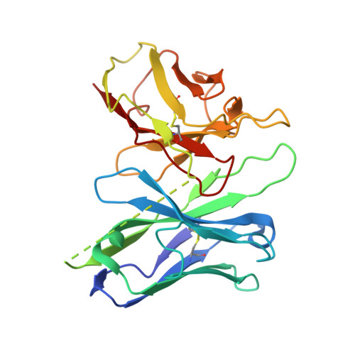
Cryo-EM structure of the human chemerin receptor 1-Gi protein complex bound to the C-terminal nonapeptide of chemerin.

Wang, J.Chen, G.Liao, Q.Lyu, W.Liu, A.Zhu, L.Du, Y.Ye, R.D.

(2023) Proc Natl Acad Sci U S A 120: e2214324120-e2214324120

  • DOI: https://doi.org/10.1073/pnas.2214324120
  • Primary Citation of Related Structures:  
    7YKD

  • PubMed Abstract: 

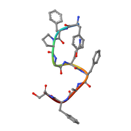
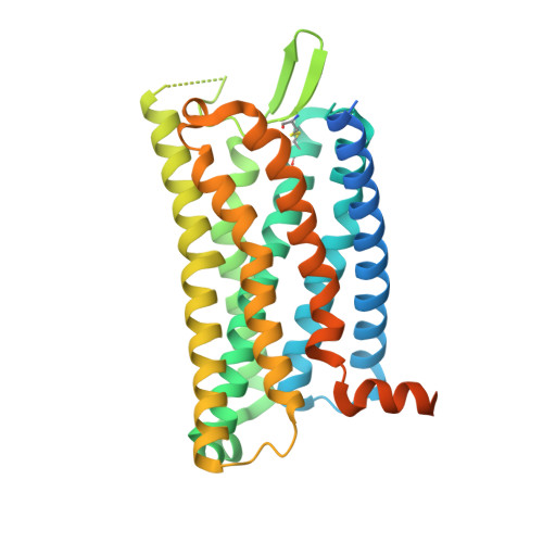
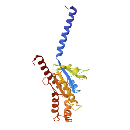
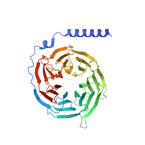
    Chemerin is a processed protein that acts on G protein-coupled receptors (GPCRs) for its chemotactic and adipokine activities. The biologically active chemerin (chemerin 21-157) results from proteolytic cleavage of prochemerin and uses its C-terminal peptide containing the sequence YFPGQFAFS for receptor activation. Here we report a high-resolution cryo-electron microscopy (cryo-EM) structure of human chemerin receptor 1 (CMKLR1) bound to the C-terminal nonapeptide of chemokine (C9) in complex with Gi proteins. C9 inserts its C terminus into the binding pocket and is stabilized through hydrophobic interactions involving its Y1, F2, F6, and F8, as well as polar interactions between G4, S9, and several amino acids lining the binding pocket of CMKLR1. Microsecond scale molecular dynamics simulations support a balanced force distribution across the whole ligand-receptor interface that enhances thermodynamic stability of the captured binding pose of C9. The C9 interaction with CMKLR1 is drastically different from chemokine recognition by chemokine receptors, which follow a two-site two-step model. In contrast, C9 takes an "S"-shaped pose in the binding pocket of CMKLR1 much like angiotensin II in the AT1 receptor. Our mutagenesis and functional analyses confirmed the cryo-EM structure and key residues in the binding pocket for these interactions. Our findings provide a structural basis for chemerin recognition by CMKLR1 for the established chemotactic and adipokine activities.


  • Organizational Affiliation

    Kobilka Institute of Innovative Drug Discovery, School of Medicine, The Chinese University of Hong Kong, Shenzhen, Guangdong 518172, P.R. China.


Macromolecules

Find similar proteins by:  Sequence   |   3D Structure  

Entity ID: 1
MoleculeChains Sequence LengthOrganismDetailsImage
Retinoic acid receptor responder protein 2A [auth L]9Homo sapiensMutation(s): 0 
Gene Names: RARRES2TIG2
Membrane Entity: Yes 
UniProt & NIH Common Fund Data Resources
Find proteins for Q99969 (Homo sapiens)
Explore Q99969 
Go to UniProtKB:  Q99969
PHAROS:  Q99969
GTEx:  ENSG00000106538 
Entity Groups  
UniProt GroupQ99969
Sequence Annotations
Expand
  • Reference Sequence
Find similar proteins by:  (by identity cutoff)  |  3D Structure
Entity ID: 2
MoleculeChains Sequence LengthOrganismDetailsImage
Chemerin-like receptor 1B [auth A]373Homo sapiensMutation(s): 0 
Gene Names: CMKLR1CHEMR23DEZ
Membrane Entity: Yes 
UniProt & NIH Common Fund Data Resources
Find proteins for Q99788 (Homo sapiens)
Explore Q99788 
Go to UniProtKB:  Q99788
PHAROS:  Q99788
GTEx:  ENSG00000174600 
Entity Groups  
Sequence Clusters30% Identity50% Identity70% Identity90% Identity95% Identity100% Identity
UniProt GroupQ99788
Sequence Annotations
Expand
  • Reference Sequence
Find similar proteins by:  (by identity cutoff)  |  3D Structure
Entity ID: 3
MoleculeChains Sequence LengthOrganismDetailsImage
Guanine nucleotide-binding protein G(i) subunit alpha-1351Homo sapiensMutation(s): 2 
Gene Names: GNAI1
Membrane Entity: Yes 
UniProt & NIH Common Fund Data Resources
Find proteins for P63096 (Homo sapiens)
Explore P63096 
Go to UniProtKB:  P63096
PHAROS:  P63096
GTEx:  ENSG00000127955 
Entity Groups  
Sequence Clusters30% Identity50% Identity70% Identity90% Identity95% Identity100% Identity
UniProt GroupP63096
Sequence Annotations
Expand
  • Reference Sequence
Find similar proteins by:  (by identity cutoff)  |  3D Structure
Entity ID: 4
MoleculeChains Sequence LengthOrganismDetailsImage
Guanine nucleotide-binding protein G(I)/G(S)/G(T) subunit beta-1D [auth B]340Homo sapiensMutation(s): 0 
Gene Names: GNB1
UniProt & NIH Common Fund Data Resources
Find proteins for P62873 (Homo sapiens)
Explore P62873 
Go to UniProtKB:  P62873
PHAROS:  P62873
GTEx:  ENSG00000078369 
Entity Groups  
Sequence Clusters30% Identity50% Identity70% Identity90% Identity95% Identity100% Identity
UniProt GroupP62873
Sequence Annotations
Expand
  • Reference Sequence
Find similar proteins by:  (by identity cutoff)  |  3D Structure
Entity ID: 5
MoleculeChains Sequence LengthOrganismDetailsImage
Guanine nucleotide-binding protein G(I)/G(S)/G(O) subunit gamma-2E [auth G]71Homo sapiensMutation(s): 0 
Gene Names: GNG2
Membrane Entity: Yes 
UniProt & NIH Common Fund Data Resources
Find proteins for P59768 (Homo sapiens)
Explore P59768 
Go to UniProtKB:  P59768
PHAROS:  P59768
GTEx:  ENSG00000186469 
Entity Groups  
Sequence Clusters30% Identity50% Identity70% Identity90% Identity95% Identity100% Identity
UniProt GroupP59768
Sequence Annotations
Expand
  • Reference Sequence
Find similar proteins by:  (by identity cutoff)  |  3D Structure
Entity ID: 6
MoleculeChains Sequence LengthOrganismDetailsImage
scFV16F [auth S]247Vicugna pacosMutation(s): 0 
Entity Groups  
Sequence Clusters30% Identity50% Identity70% Identity90% Identity95% Identity100% Identity
Sequence Annotations
Expand
  • Reference Sequence
Experimental Data & Validation

Experimental Data

  • Method: ELECTRON MICROSCOPY
  • Resolution: 2.81 Å
  • Aggregation State: PARTICLE 
  • Reconstruction Method: SINGLE PARTICLE 

Structure Validation

View Full Validation Report


Entry History & Funding Information

Deposition Data


Funding OrganizationLocationGrant Number
Not funded--

Revision History  (Full details and data files)

  • Version 1.0: 2023-04-19
    Type: Initial release