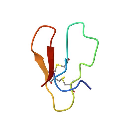
Structural insight into why S-linked glycosylation cannot adequately mimic the role of natural O-glycosylation.
Chen, C., Ma, B., Wang, Y., Cui, Q., Yao, L., Li, Y., Chen, B., Feng, Y., Tan, Z.(2023) Int J Biol Macromol 253: 126649-126649
- PubMed: 37666405 Search on PubMed
- DOI: https://doi.org/10.1016/j.ijbiomac.2023.126649
- Primary Citation Related Structures:
7YHF, 7YHG, 7YHH, 7YHI - PubMed Abstract:
There is an increasing interest in using S-glycosylation as a replacement for the more commonly occurring O-glycosylation, aiming to enhance the resistance of glycans against chemical hydrolysis and enzymatic degradation. However, previous studies have demonstrated that these two types of glycosylation exert distinct effects on protein properties and functions. In order to elucidate the structural basis behind the observed differences, we conducted a systematic and comparative analysis of 6 differently glycosylated forms of a model glycoprotein, CBM, using NMR spectroscopy and molecular dynamic simulations. Our findings revealed that the different stabilizing effects of S- and O-glycosylation could be attributed to altered hydrogen-bonding capability between the glycan and the polypeptide chain, and their diverse impacts on binding affinity could be elucidated by examining the interactions and motion dynamics of glycans in substrate-bound states. Overall, this study underscores the pivotal role of the glycosidic linkage in shaping the function of glycosylation and advises caution when switching glycosylation types in protein glycoengineering.
- CAS Key Laboratory of Biofuels, Shandong Provincial Key Laboratory of Synthetic Biology, Qingdao Institute of Bioenergy and Bioprocess Technology, Chinese Academy of Sciences, Qingdao 266101, China; Shandong Engineering Laboratory of Single Cell Oil, Qingdao Institute of Bioenergy and Bioprocess Technology, Chinese Academy of Sciences, Qingdao 266101, China; Shandong Energy Institute, Qingdao, Shandong 266101, China; Qingdao New Energy Shandong Laboratory, Qingdao, Shandong 266101, China; University of Chinese Academy of Sciences, Beijing 100049, China.
Organizational Affiliation: