Structural and biochemical insights into Zn 2+ -bound EF-hand proteins, EFhd1 and EFhd2.
Mun, S.A., Park, J., Kang, J.Y., Park, T., Jin, M., Yang, J., Eom, S.H.(2023) IUCrJ 10: 233-245
- PubMed: 36862489 Search on PubMedSearch on PubMed Central
- DOI: https://doi.org/10.1107/S2052252523001501
- Primary Citation Related Structures:
7YGV, 7YGW, 7YGY - PubMed Abstract:
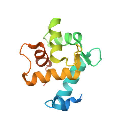
EF-hand proteins, which contain a Ca 2+ -binding EF-hand motif, are involved in regulating diverse cellular functions. Ca 2+ binding induces conformational changes that modulate the activities of EF-hand proteins. Moreover, these proteins occasionally modify their activities by coordinating metals other than Ca 2+ , including Mg 2+ , Pb 2+ and Zn 2+ , within their EF-hands. EFhd1 and EFhd2 are homologous EF-hand proteins with similar structures. Although separately localized within cells, both are actin-binding proteins that modulate F-actin rearrangement through Ca 2+ -independent actin-binding and Ca 2+ -dependent actin-bundling activity. Although Ca 2+ is known to affect the activities of EFhd1 and EFhd2, it is not known whether their actin-related activities are affected by other metals. Here, the crystal structures of the EFhd1 and EFhd2 core domains coordinating Zn 2+ ions within their EF-hands are reported. The presence of Zn 2+ within EFhd1 and EFhd2 was confirmed by analyzing anomalous signals and the difference between anomalous signals using data collected at the peak positions as well as low-energy remote positions at the Zn K-edge. EFhd1 and EFhd2 were also found to exhibit Zn 2+ -independent actin-binding and Zn 2+ -dependent actin-bundling activity. This suggests the actin-related activities of EFhd1 and EFhd2 could be regulated by Zn 2+ as well as Ca 2+ .
- School of Life Sciences, Gwangju Institute of Science and Technology, Gwangju, Republic of Korea.
Organizational Affiliation: