7YAG

CryoEM structure of SPCA1a in E1-Ca-AMPPCP state subclass 1


Experimental Data Snapshot

  • Method: ELECTRON MICROSCOPY
  • Resolution: 3.10 Å
  • Aggregation State: PARTICLE 
  • Reconstruction Method: SINGLE PARTICLE 

wwPDB Validation   3D Report Full Report


This is version 1.0 of the entry. See complete history


Literature

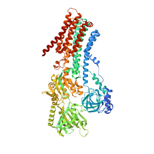
Cryo-EM structures of human SPCA1a reveal the mechanism of Ca 2+ /Mn 2+ transport into the Golgi apparatus.

Chen, Z.Watanabe, S.Hashida, H.Inoue, M.Daigaku, Y.Kikkawa, M.Inaba, K.

(2023) Sci Adv 9: eadd9742-eadd9742

  • DOI: https://doi.org/10.1126/sciadv.add9742
  • Primary Citation of Related Structures:  
    7YAG, 7YAH, 7YAI, 7YAJ, 7YAM

  • PubMed Abstract: 

    Secretory pathway Ca 2+ /Mn 2+ ATPase 1 (SPCA1) actively transports cytosolic Ca 2+ and Mn 2+ into the Golgi lumen, playing a crucial role in cellular calcium and manganese homeostasis. Detrimental mutations of the ATP2C1 gene encoding SPCA1 cause Hailey-Hailey disease. Here, using nanobody/megabody technologies, we determined cryo-electron microscopy structures of human SPCA1a in the ATP and Ca 2+ /Mn 2+ -bound (E1-ATP) state and the metal-free phosphorylated (E2P) state at 3.1- to 3.3-Å resolutions. The structures revealed that Ca 2+ and Mn 2+ share the same metal ion-binding pocket with similar but notably different coordination geometries in the transmembrane domain, corresponding to the second Ca 2+ -binding site in sarco/endoplasmic reticulum Ca 2+ -ATPase (SERCA). In the E1-ATP to E2P transition, SPCA1a undergoes similar domain rearrangements to those of SERCA. Meanwhile, SPCA1a shows larger conformational and positional flexibility of the second and sixth transmembrane helices, possibly explaining its wider metal ion specificity. These structural findings illuminate the unique mechanisms of SPCA1a-mediated Ca 2+ /Mn 2+ transport.


  • Organizational Affiliation

    Institute of Multidisciplinary Research for Advanced Materials, Tohoku University, Sendai, Miyagi 980-8577, Japan.


Macromolecules
Find similar proteins by:  (by identity cutoff)  |  3D Structure
Entity ID: 1
MoleculeChains Sequence LengthOrganismDetailsImage
Calcium-transporting ATPase type 2C member 1947Homo sapiensMutation(s): 0 
Gene Names: ATP2C1KIAA1347PMR1LHUSSY-28
EC: 7.2.2.10
Membrane Entity: Yes 
UniProt & NIH Common Fund Data Resources
Find proteins for P98194 (Homo sapiens)
Explore P98194 
Go to UniProtKB:  P98194
PHAROS:  P98194
GTEx:  ENSG00000017260 
Entity Groups  
Sequence Clusters30% Identity50% Identity70% Identity90% Identity95% Identity100% Identity
UniProt GroupP98194
Sequence Annotations
Expand
  • Reference Sequence
Find similar proteins by:  (by identity cutoff)  |  3D Structure
Entity ID: 2
MoleculeChains Sequence LengthOrganismDetailsImage
nanobody head piece of megabodyB [auth C]128Vicugna pacosMutation(s): 0 
Entity Groups  
Sequence Clusters30% Identity50% Identity70% Identity90% Identity95% Identity100% Identity
Sequence Annotations
Expand
  • Reference Sequence
Small Molecules
Experimental Data & Validation

Experimental Data

  • Method: ELECTRON MICROSCOPY
  • Resolution: 3.10 Å
  • Aggregation State: PARTICLE 
  • Reconstruction Method: SINGLE PARTICLE 

Structure Validation

View Full Validation Report


Entry History & Funding Information

Deposition Data


Funding OrganizationLocationGrant Number
Japan Agency for Medical Research and Development (AMED)Japan21gm1410006h0001, JP19am0101115
Japan Society for the Promotion of Science (JSPS)Japan18H03978, 21H04758, 21H05247, 21K15036

Revision History  (Full details and data files)

  • Version 1.0: 2023-03-22
    Type: Initial release