7YA2 | pdb_00007ya2

Crystal structure of capsular polysaccharide synthesis enzyme CapG from Staphylococcus aureus


Experimental Data Snapshot

  • Method: X-RAY DIFFRACTION
  • Resolution: 3.20 Å
  • R-Value Free: 
    0.241 (Depositor), 0.240 (DCC) 
  • R-Value Work: 
    0.183 (Depositor), 0.185 (DCC) 

Starting Model: experimental
View more details

wwPDB Validation 3D Report Full Report

Validation slider image for 7YA2

This is version 1.1 of the entry. See complete history

Literature

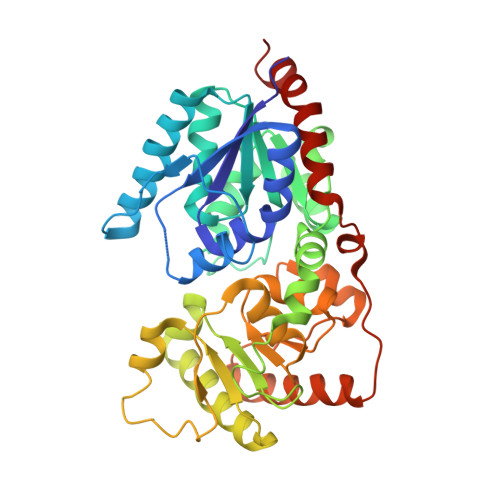
Crystal structure of the capsular polysaccharide-synthesis enzyme CapG from Staphylococcus aureus.

Tien, N.Ho, C.Y.Lai, S.J.Lin, Y.C.Yang, C.S.Wang, Y.C.Huang, W.C.Chen, Y.Chang, J.J.

(2022) Acta Crystallogr F Struct Biol Commun 78: 378-385

  • DOI: https://doi.org/10.1107/S2053230X22008743
  • Primary Citation Related Structures: 
    7YA2

  • PubMed Abstract: 

    Bacterial capsular polysaccharides provide protection against environmental stress and immune evasion from the host immune system, and are therefore considered to be attractive therapeutic targets for the development of anti-infectious reagents. Here, we focused on CapG, one of the key enzymes in the synthesis pathway of capsular polysaccharides type 5 (CP5) from the opportunistic pathogen Staphylococcus aureus. SaCapG catalyses the 2-epimerization of UDP-N-acetyl-D-talosamine (UDP-TalNAc) to UDP-N-acetyl-D-fucosamine (UDP-FucNAc), which is one of the nucleotide-activated precursors for the synthesis of the trisaccharide repeating units of CP5. Here, the cloning, expression and purification of recombinant SaCapG are reported. After extensive efforts, single crystals of SaCapG were successfully obtained which belonged to space group C2 and exhibited unit-cell parameters a = 302.91, b = 84.34, c = 145.09 Å, β = 110.65°. The structure was solved by molecular replacement and was refined to 3.2 Å resolution. The asymmetric unit revealed a homohexameric assembly of SaCapG, which was consistent with gel-filtration analysis. Structural comparison with UDP-N-acetyl-D-glucosamine 2-epimerase from Methanocaldococcus jannaschii identified α2, the α2-α3 loop and α10 as a gate-regulated switch controlling substrate entry and/or product release.


  • Organizational Affiliation
    • Department of Laboratory Medicine, China Medical University Hospital, Taichung, Taiwan.

Macromolecule Content 

  • Total Structure Weight: 257.43 kDa 
  • Atom Count: 18,050 
  • Modeled Residue Count: 2,242 
  • Deposited Residue Count: 2,244 
  • Unique protein chains: 1

Macromolecules

Find similar proteins by:|  3D Structure
Entity ID: 1
MoleculeChains  Sequence LengthOrganismDetailsImage
Capsular polysaccharide synthesis enzyme CapG
A, B, C, D, E
A, B, C, D, E, F
374Staphylococcus aureus subsp. aureus str. NewmanMutation(s): 0 
Gene Names: capGNWMN_0101CNH35_00585
UniProt
Find proteins for A0A0H3K5M7 (Staphylococcus aureus (strain Newman))
Explore A0A0H3K5M7 
Go to UniProtKB:  A0A0H3K5M7
Entity Groups
Sequence Clusters30% Identity50% Identity70% Identity90% Identity95% Identity100% Identity
UniProt GroupA0A0H3K5M7
Sequence Annotations
Expand
Reference Sequence

Experimental Data & Validation

Experimental Data

  • Method: X-RAY DIFFRACTION
  • Resolution: 3.20 Å
  • R-Value Free:  0.241 (Depositor), 0.240 (DCC) 
  • R-Value Work:  0.183 (Depositor), 0.185 (DCC) 
Space Group: C 1 2 1
Unit Cell:
Length ( Å )Angle ( ˚ )
a = 302.909α = 90
b = 84.34β = 110.65
c = 145.085γ = 90
Software Package:
Software NamePurpose
REFMACrefinement
HKL-2000data reduction
HKL-2000data scaling
MOLREPphasing

Structure Validation

View Full Validation Report


Entry History 

& Funding Information

Deposition Data


Funding OrganizationLocationGrant Number
Ministry of Science and Technology (MoST, Taiwan)Taiwan109-2311-B-241-001

Revision History  (Full details and data files)

  • Version 1.0: 2022-11-16
    Type: Initial release
  • Version 1.1: 2023-11-29
    Changes: Data collection, Refinement description