Characterization of structure of peptidyl-tRNA hydrolase from Enterococcus faecium and its inhibition by a pyrrolinone compound.
Pandey, R., Kaul, G., Akhir, A., Saxena, D., Shukla, M., Mundra, S., Zohib, M., Singh, S., Pal, R.K., Tripathi, S., Jain, A., Chopra, S., Arora, A.(2024) Int J Biol Macromol 275: 133445-133445
- PubMed: 38945334 Search on PubMed
- DOI: https://doi.org/10.1016/j.ijbiomac.2024.133445
- Primary Citation Related Structures:
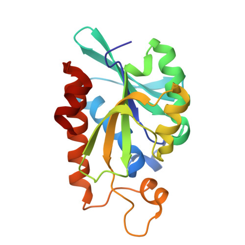
7Y52, 9LS8 - PubMed Abstract:
In bacteria, peptidyl-tRNA hydrolase (Pth, E.C. 3.1.1.29) is a ubiquitous and essential enzyme for preventing the accumulation of peptidyl-tRNA and sequestration of tRNA. Pth is an esterase that cleaves the ester bond between peptide and tRNA. Here, we present the crystal structure of Pth from Enterococcus faecium (EfPth) at a resolution of 1.92 Å. The two molecules in the asymmetric unit differ in the orientation of sidechain of N66, a conserved residue of the catalytic site. Enzymatic hydrolysis of substrate α-N-BODIPY-lysyl-tRNA Lys (BLT) by EfPth was characterized by Michaelis-Menten parameters K M 163.5 nM and Vmax 1.9 nM/s. Compounds having pyrrolinone scaffold were tested for inhibition of Pth and one compound, 1040-C, was found to have IC 50 of 180 nM. Antimicrobial activity profiling was done for 1040-C. It exhibited equipotent activity against drug-susceptible and resistant S. aureus (MRSA and VRSA) and Enterococcus (VSE and VRE) with MICs 2-8 μg/mL. 1040-C synergized with gentamicin and the combination was effective against the gentamicin resistant S. aureus strain NRS-119. 1040-C was found to reduce biofilm mass of S. aureus to an extent similar to Vancomycin. In a murine model of infection, 1040-C was able to reduce bacterial load to an extent comparable to Vancomycin.
- Biochemistry and Structural Biology Division, CSIR - Central Drug Research Institute, Lucknow 226031, India; Academy of Scientific and Innovative Research (AcSIR), Ghaziabad 201002, India.
Organizational Affiliation: