7Y3N

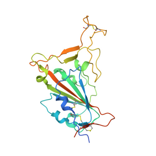
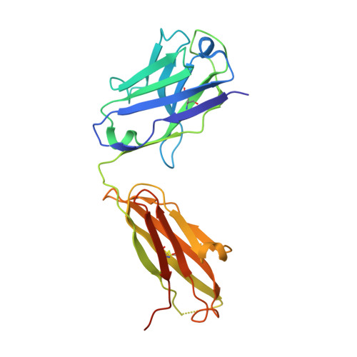
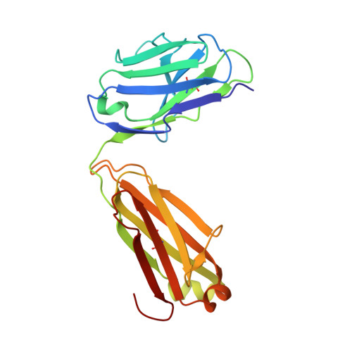
Crystal structure of SARS-CoV receptor binding domain in complex with human antibody BIOLS56


Experimental Data Snapshot

  • Method: X-RAY DIFFRACTION
  • Resolution: 2.97 Å
  • R-Value Free: 0.263 
  • R-Value Work: 0.220 
  • R-Value Observed: 0.223 

wwPDB Validation   3D Report Full Report


Ligand Structure Quality Assessment 


This is version 1.0 of the entry. See complete history


Literature

Defining a de novo non-RBM antibody as RBD-8 and its synergistic rescue of immune-evaded antibodies to neutralize Omicron SARS-CoV-2.

Rao, X.Zhao, R.Tong, Z.Guo, S.Peng, W.Liu, K.Li, S.Wu, L.Tong, J.Chai, Y.Han, P.Wang, F.Jia, P.Li, Z.Zhao, X.Li, D.Zhang, R.Zhang, X.Zou, W.Li, W.Wang, Q.Gao, G.F.Wu, Y.Dai, L.Gao, F.

(2023) Proc Natl Acad Sci U S A 120: e2314193120-e2314193120

  • DOI: https://doi.org/10.1073/pnas.2314193120
  • Primary Citation of Related Structures:  
    7Y3N, 7Y3O, 8HRD

  • PubMed Abstract: 

    Currently, monoclonal antibodies (MAbs) targeting the SARS-CoV-2 receptor binding domain (RBD) of spike (S) protein are classified into seven classes based on their binding epitopes. However, most of these antibodies are seriously impaired by SARS-CoV-2 Omicron and its subvariants, especially the recent BQ.1.1, XBB and its derivatives. Identification of broadly neutralizing MAbs against currently circulating variants is imperative. In this study, we identified a "breathing" cryptic epitope in the S protein, named as RBD-8. Two human MAbs, BIOLS56 and IMCAS74, were isolated recognizing this epitope with broad neutralization abilities against tested sarbecoviruses, including SARS-CoV, pangolin-origin coronaviruses, and all the SARS-CoV-2 variants tested (Omicron BA.4/BA.5, BQ.1.1, and XBB subvariants). Searching through the literature, some more RBD-8 MAbs were defined. More importantly, BIOLS56 rescues the immune-evaded antibody, RBD-5 MAb IMCAS-L4.65, by making a bispecific MAb, to neutralize BQ.1 and BQ.1.1, thereby producing an MAb to cover all the currently circulating Omicron subvariants. Structural analysis reveals that the neutralization effect of RBD-8 antibodies depends on the extent of epitope exposure, which is affected by the angle of antibody binding and the number of up-RBDs induced by angiotensin-converting enzyme 2 binding. This cryptic epitope which recognizes non- receptor binding motif (non-RBM) provides guidance for the development of universal therapeutic antibodies and vaccines against COVID-19.


  • Organizational Affiliation

    Laboratory of Protein Engineering and Vaccines, Tianjin Institute of Industrial Biotechnology, Chinese Academy of Sciences, Tianjin 300308, China.


Macromolecules
Find similar proteins by:  (by identity cutoff)  |  3D Structure
Entity ID: 1
MoleculeChains Sequence LengthOrganismDetailsImage
Spike protein S1A,
D [auth B],
G [auth E]
228Severe acute respiratory syndrome-related coronavirusMutation(s): 0 
Gene Names: S2
UniProt
Find proteins for P59594 (Severe acute respiratory syndrome coronavirus)
Explore P59594 
Go to UniProtKB:  P59594
Entity Groups  
Sequence Clusters30% Identity50% Identity70% Identity90% Identity95% Identity100% Identity
UniProt GroupP59594
Sequence Annotations
Expand
  • Reference Sequence
Find similar proteins by:  (by identity cutoff)  |  3D Structure
Entity ID: 2
MoleculeChains Sequence LengthOrganismDetailsImage
Heavy chain of BIOLS56B [auth H],
E [auth C],
H [auth F]
237Homo sapiensMutation(s): 0 
Entity Groups  
Sequence Clusters30% Identity50% Identity70% Identity90% Identity95% Identity100% Identity
Sequence Annotations
Expand
  • Reference Sequence
Find similar proteins by:  (by identity cutoff)  |  3D Structure
Entity ID: 3
MoleculeChains Sequence LengthOrganismDetailsImage
Light chain of BIOLS56C [auth L],
F [auth D],
I [auth G]
215Homo sapiensMutation(s): 0 
Entity Groups  
Sequence Clusters30% Identity50% Identity70% Identity90% Identity95% Identity100% Identity
Sequence Annotations
Expand
  • Reference Sequence
Oligosaccharides

Help

Entity ID: 4
MoleculeChains Length2D Diagram Glycosylation3D Interactions
2-acetamido-2-deoxy-beta-D-glucopyranose-(1-4)-[alpha-L-fucopyranose-(1-6)]2-acetamido-2-deoxy-beta-D-glucopyranoseJ [auth I]3N-Glycosylation
Glycosylation Resources
GlyTouCan:  G21290RB
GlyCosmos:  G21290RB
GlyGen:  G21290RB
Small Molecules
Ligands 1 Unique
IDChains Name / Formula / InChI Key2D Diagram3D Interactions
NAG
Query on NAG

Download Ideal Coordinates CCD File 
K [auth B]2-acetamido-2-deoxy-beta-D-glucopyranose
C8 H15 N O6
OVRNDRQMDRJTHS-FMDGEEDCSA-N
Experimental Data & Validation

Experimental Data

  • Method: X-RAY DIFFRACTION
  • Resolution: 2.97 Å
  • R-Value Free: 0.263 
  • R-Value Work: 0.220 
  • R-Value Observed: 0.223 
  • Space Group: P 21 21 21
Unit Cell:
Length ( Å )Angle ( ˚ )
a = 112.483α = 90
b = 159.018β = 90
c = 175.99γ = 90
Software Package:
Software NamePurpose
HKL-2000data reduction
HKL-2000data scaling
PHENIXrefinement
PHASERphasing

Structure Validation

View Full Validation Report


Ligand Structure Quality Assessment 


Entry History & Funding Information

Deposition Data


Funding OrganizationLocationGrant Number
Chinese Academy of SciencesChinaXDB29010202

Revision History  (Full details and data files)

  • Version 1.0: 2023-12-27
    Type: Initial release