7Y1R | pdb_00007y1r

Human L-TGF-beta1 in complex with the anchor protein LRRC33


Experimental Data Snapshot

  • Method: ELECTRON MICROSCOPY
  • Resolution: 4.01 Å
  • Aggregation State: PARTICLE 
  • Reconstruction Method: SINGLE PARTICLE 

wwPDB Validation 3D Report Full Report

Validation slider image for 7Y1R

This is version 1.2 of the entry. See complete history. 

Literature

Specificity of TGF-beta 1 signal designated by LRRC33 and integrin alpha V beta 8.

Duan, Z., Lin, X., Wang, L., Zhen, Q., Jiang, Y., Chen, C., Yang, J., Lee, C.H., Qin, Y., Li, Y., Zhao, B., Wang, J., Zhang, Z.

(2022) Nat Commun 13: 4988-4988

  • DOI: https://doi.org/10.1038/s41467-022-32655-9
  • Primary Citation Related Structures: 
    7Y1R, 7Y1T

  • PubMed Abstract: 

    Myeloid lineage cells present the latent form of transforming growth factor-β1 (L-TGF-β1) to the membrane using an anchor protein LRRC33. Integrin α V β 8 activates extracellular L-TGF-β1 to trigger the downstream signaling functions. However, the mechanism designating the specificity of TGF-β1 presentation and activation remains incompletely understood. Here, we report cryo-EM structures of human L-TGF-β1/LRRC33 and integrin α V β 8 /L-TGF-β1 complexes. Combined with biochemical and cell-based analyses, we demonstrate that LRRC33 only presents L-TGF-β1 but not the -β2 or -β3 isoforms due to difference of key residues on the growth factor domains. Moreover, we reveal a 2:2 binding mode of integrin α V β 8 and L-TGF-β1, which shows higher avidity and more efficient L-TGF-β1 activation than previously reported 1:2 binding mode. We also uncover that the disulfide-linked loop of the integrin subunit β 8 determines its exquisite affinity to L-TGF-β1. Together, our findings provide important insights into the specificity of TGF-β1 signaling achieved by LRRC33 and integrin α V β 8 .


  • Organizational Affiliation: 
    • State Key Laboratory of Membrane Biology, Center for Life Sciences, School of Life Sciences, Peking University, 100871, Beijing, China.

Macromolecule Content 

  • Total Structure Weight: 162.21 kDa 
  • Atom Count: 8,565 
  • Modeled Residue Count: 1,041 
  • Deposited Residue Count: 1,410 
  • Unique protein chains: 2

Macromolecules

Find similar proteins by:|  3D Structure
Entity ID: 1
MoleculeChains  Sequence LengthOrganismDetailsImage
Transforming growth factor beta-1 proprotein
A, B
377Homo sapiensMutation(s): 0 
Gene Names: TGFB1
UniProt & NIH Common Fund Data Resources
Find proteins for P01137 (Homo sapiens)
Explore P01137 
Go to UniProtKB:  P01137
GTEx:  ENSG00000105329 
Entity Groups
Sequence Clusters30% Identity50% Identity70% Identity90% Identity95% Identity100% Identity
UniProt GroupP01137
Glycosylation
Glycosylation Sites: 2Go to GlyGen: P01137-1
Sequence Annotations
Expand
Reference Sequence
Find similar proteins by:|  3D Structure
Entity ID: 2
MoleculeChains  Sequence LengthOrganismDetailsImage
Transforming growth factor beta activator LRRC33C [auth E]656Homo sapiensMutation(s): 0 
Gene Names: LRRC33
UniProt & NIH Common Fund Data Resources
Find proteins for Q86YC3 (Homo sapiens)
Explore Q86YC3 
Go to UniProtKB:  Q86YC3
PHAROS:  Q86YC3
GTEx:  ENSG00000174004 
Entity Groups
Sequence Clusters30% Identity50% Identity70% Identity90% Identity95% Identity100% Identity
UniProt GroupQ86YC3
Glycosylation
Glycosylation Sites: 4Go to GlyGen: Q86YC3-1
Sequence Annotations
Expand
Reference Sequence

Oligosaccharides

Help  
Entity ID: 3
MoleculeChains Length2D Diagram GlycosylationD Interactions
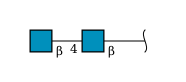
beta-D-mannopyranose-(1-4)-2-acetamido-2-deoxy-beta-D-glucopyranose-(1-4)-2-acetamido-2-deoxy-beta-D-glucopyranoseD [auth L],
G [auth F],
H [auth G],
I [auth H],
J [auth I]
3N-Glycosylation
Glycosylation Resources
GlyTouCan: G15407YE
GlyCosmos: G15407YE
GlyGen: G15407YE
Entity ID: 4
MoleculeChains Length2D Diagram GlycosylationD Interactions
2-acetamido-2-deoxy-beta-D-glucopyranose-(1-4)-2-acetamido-2-deoxy-beta-D-glucopyranoseE [auth C]2N-Glycosylation
Glycosylation Resources
GlyTouCan: G42666HT
GlyCosmos: G42666HT
GlyGen: G42666HT
Entity ID: 5
MoleculeChains Length2D Diagram GlycosylationD Interactions
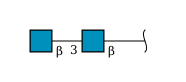
2-acetamido-2-deoxy-beta-D-glucopyranose-(1-3)-2-acetamido-2-deoxy-beta-D-glucopyranoseF [auth D]2N-Glycosylation
Glycosylation Resources
GlyTouCan: G79924RM
GlyCosmos: G79924RM
GlyGen: G79924RM

Small Molecules

Ligands 1 Unique
IDChains Name / Formula / InChI Key2D Diagram3D Interactions
NAG
(Subject of Investigation/LOI)

Query on NAG


Download:Ideal Coordinates CCD File
K [auth B]2-acetamido-2-deoxy-beta-D-glucopyranose
C8 H15 N O6
OVRNDRQMDRJTHS-FMDGEEDCSA-N

Experimental Data & Validation

Experimental Data

  • Method: ELECTRON MICROSCOPY
  • Resolution: 4.01 Å
  • Aggregation State: PARTICLE 
  • Reconstruction Method: SINGLE PARTICLE 

Structure Validation

View Full Validation Report


Entry History 

& Funding Information

Deposition Data


Funding OrganizationLocationGrant Number
National Natural Science Foundation of China (NSFC)China32171201

Revision History  (Full details and data files)

  • Version 1.0: 2022-08-31
    Type: Initial release
  • Version 1.1: 2022-09-07
    Changes: Database references
  • Version 1.2: 2024-10-23
    Changes: Data collection, Structure summary