7Y1A

Lateral hexamer


Experimental Data Snapshot

  • Method: ELECTRON MICROSCOPY
  • Resolution: 6.30 Å
  • Aggregation State: CELL 
  • Reconstruction Method: SINGLE PARTICLE 

wwPDB Validation   3D Report Full Report


This is version 1.2 of the entry. See complete history


Literature

In situ structure of the red algal phycobilisome-PSII-PSI-LHC megacomplex.

You, X.Zhang, X.Cheng, J.Xiao, Y.Ma, J.Sun, S.Zhang, X.Wang, H.W.Sui, S.F.

(2023) Nature 616: 199-206

  • DOI: https://doi.org/10.1038/s41586-023-05831-0
  • Primary Citation of Related Structures:  
    7Y1A, 7Y4L, 7Y5E, 7Y7A

  • PubMed Abstract: 

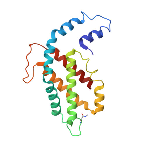
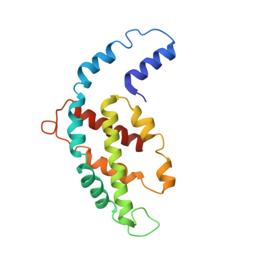
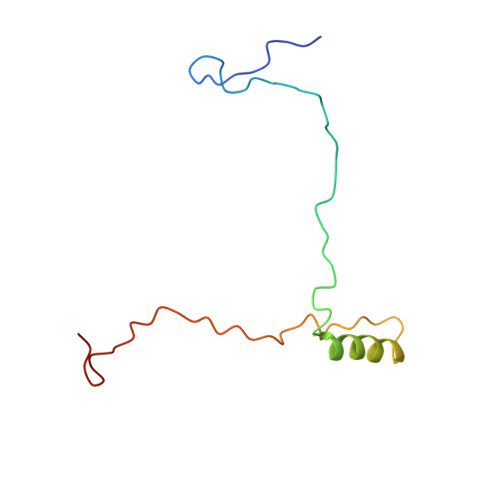
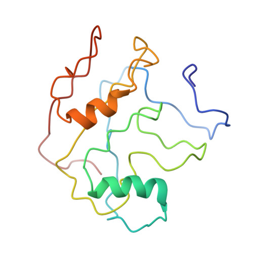
    In oxygenic photosynthetic organisms, light energy is captured by antenna systems and transferred to photosystem II (PSII) and photosystem I (PSI) to drive photosynthesis 1,2 . The antenna systems of red algae consist of soluble phycobilisomes (PBSs) and transmembrane light-harvesting complexes (LHCs) 3 . Excitation energy transfer pathways from PBS to photosystems remain unclear owing to the lack of structural information. Here we present in situ structures of PBS-PSII-PSI-LHC megacomplexes from the red alga Porphyridium purpureum at near-atomic resolution using cryogenic electron tomography and in situ single-particle analysis 4 , providing interaction details between PBS, PSII and PSI. The structures reveal several unidentified and incomplete proteins and their roles in the assembly of the megacomplex, as well as a huge and sophisticated pigment network. This work provides a solid structural basis for unravelling the mechanisms of PBS-PSII-PSI-LHC megacomplex assembly, efficient energy transfer from PBS to the two photosystems, and regulation of energy distribution between PSII and PSI.


  • Organizational Affiliation

    State Key Laboratory of Membrane Biology, Beijing Frontier Research Center for Biological Structures, Beijing Advanced Innovation Center for Structural Biology, School of Life Sciences, Tsinghua University, Beijing, China.


Macromolecules
Find similar proteins by:  (by identity cutoff)  |  3D Structure
Entity ID: 1
MoleculeChains Sequence LengthOrganismDetailsImage
B-phycoerythrin beta chain177Porphyridium purpureumMutation(s): 0 
Gene Names: cpeB
Membrane Entity: Yes 
UniProt
Find proteins for P11393 (Porphyridium purpureum)
Explore P11393 
Go to UniProtKB:  P11393
Entity Groups  
Sequence Clusters30% Identity50% Identity70% Identity90% Identity95% Identity100% Identity
UniProt GroupP11393
Sequence Annotations
Expand
  • Reference Sequence
Find similar proteins by:  (by identity cutoff)  |  3D Structure
Entity ID: 2
MoleculeChains Sequence LengthOrganismDetailsImage
Phycoerythrin alpha subunit164Porphyridium purpureumMutation(s): 0 
Gene Names: peAcpeA
Membrane Entity: Yes 
UniProt
Find proteins for P11392 (Porphyridium purpureum)
Explore P11392 
Go to UniProtKB:  P11392
Entity Groups  
Sequence Clusters30% Identity50% Identity70% Identity90% Identity95% Identity100% Identity
UniProt GroupP11392
Sequence Annotations
Expand
  • Reference Sequence
Find similar proteins by:  (by identity cutoff)  |  3D Structure
Entity ID: 3
MoleculeChains Sequence LengthOrganismDetailsImage
LRHM [auth a]87Porphyridium purpureumMutation(s): 0 
Sequence Annotations
Expand
  • Reference Sequence
Find similar proteins by:  (by identity cutoff)  |  3D Structure
Entity ID: 4
MoleculeChains Sequence LengthOrganismDetailsImage
LRHN [auth A]150Porphyridium purpureumMutation(s): 0 
Sequence Annotations
Expand
  • Reference Sequence
Small Molecules
Ligands 1 Unique
IDChains Name / Formula / InChI Key2D Diagram3D Interactions
PEB (Subject of Investigation/LOI)
Query on PEB

Download Ideal Coordinates CCD File 
AA [auth q]
BA [auth r]
CA [auth r]
DA [auth s]
EA [auth s]
AA [auth q],
BA [auth r],
CA [auth r],
DA [auth s],
EA [auth s],
FA [auth s],
GA [auth t],
HA [auth t],
IA [auth u],
JA [auth u],
KA [auth u],
LA [auth v],
MA [auth v],
NA [auth w],
O [auth y],
OA [auth w],
P [auth y],
PA [auth w],
Q [auth y],
QA [auth w],
R [auth z],
RA [auth x],
S [auth z],
SA [auth x],
T [auth 1],
U [auth 1],
V [auth 1],
W [auth p],
X [auth p],
Y [auth q],
Z [auth q]
PHYCOERYTHROBILIN
C33 H40 N4 O6
NKCBCVIFPXGHAV-WAVSMFBNSA-N
Modified Residues  1 Unique
IDChains TypeFormula2D DiagramParent
MEN
Query on MEN
A [auth y]
C [auth 1]
E [auth q]
G [auth s]
I [auth u]
A [auth y],
C [auth 1],
E [auth q],
G [auth s],
I [auth u],
K [auth w]
L-PEPTIDE LINKINGC5 H10 N2 O3ASN
Experimental Data & Validation

Experimental Data

  • Method: ELECTRON MICROSCOPY
  • Resolution: 6.30 Å
  • Aggregation State: CELL 
  • Reconstruction Method: SINGLE PARTICLE 

Structure Validation

View Full Validation Report


Entry History & Funding Information

Deposition Data


Funding OrganizationLocationGrant Number
Not funded--

Revision History  (Full details and data files)

  • Version 1.0: 2023-01-18
    Type: Initial release
  • Version 1.1: 2023-03-29
    Changes: Database references
  • Version 1.2: 2023-04-19
    Changes: Database references