7XZZ

Cryo-EM structure of the nucleosome in complex with p53


Experimental Data Snapshot

  • Method: ELECTRON MICROSCOPY
  • Resolution: 4.07 Å
  • Aggregation State: PARTICLE 
  • Reconstruction Method: SINGLE PARTICLE 

wwPDB Validation   3D Report Full Report


This is version 1.1 of the entry. See complete history


Literature

Structural basis for p53 binding to its nucleosomal target DNA sequence.

Nishimura, M.Takizawa, Y.Nozawa, K.Kurumizaka, H.

(2022) PNAS Nexus 1: pgac177-pgac177

  • DOI: https://doi.org/10.1093/pnasnexus/pgac177

  • PubMed Abstract: 

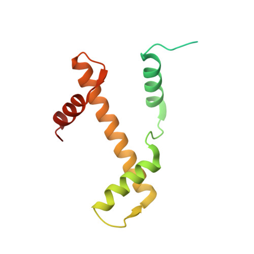
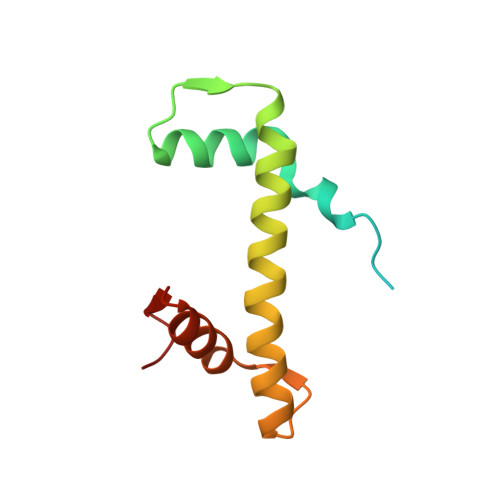
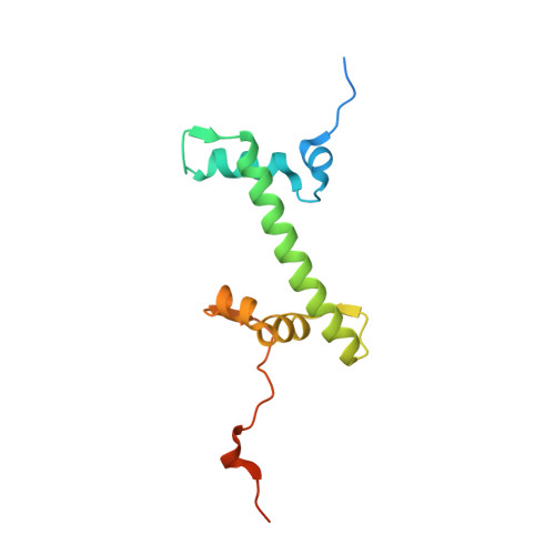
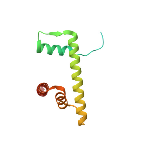
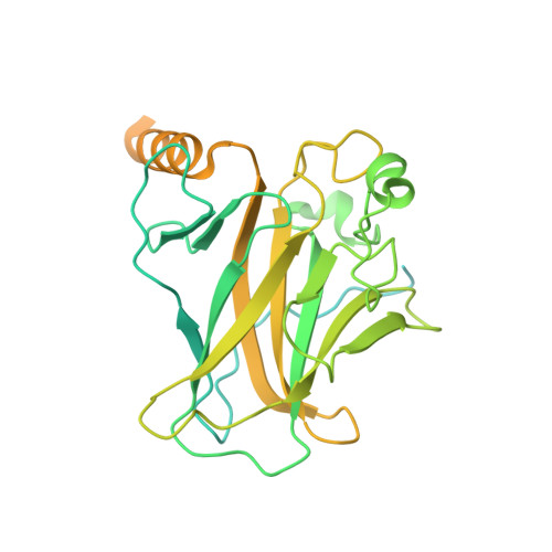
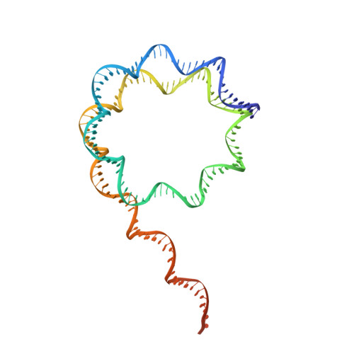
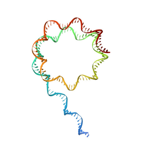
    The tumor suppressor p53 functions as a pioneer transcription factor that binds a nucleosomal target DNA sequence. However, the mechanism by which p53 binds to its target DNA in the nucleosome remains elusive. Here we report the cryo-electron microscopy structures of the p53 DNA-binding domain and the full-length p53 protein complexed with a nucleosome containing the 20 base-pair target DNA sequence of p53 (p53BS). In the p53-nucleosome structures, the p53 DNA-binding domain forms a tetramer and specifically binds to the p53BS DNA, located near the entry/exit region of the nucleosome. The nucleosomal position of the p53BS DNA is within the genomic p21 promoter region. The p53 binding peels the DNA from the histone surface, and drastically changes the DNA path around the p53BS on the nucleosome. The C-terminal domain of p53 also binds to the DNA around the center and linker DNA regions of the nucleosome, as revealed by hydroxyl radical footprinting. These results provide important structural information for understanding the mechanism by which p53 binds the nucleosome and changes the chromatin structure for gene activation.


  • Organizational Affiliation

    Laboratory of Chromatin Structure and Function, Institute for Quantitative Biosciences, The University of Tokyo, 1-1-1 Yayoi, Bunkyo-ku, Tokyo 113-0032, Japan.


Macromolecules

Find similar proteins by:  (by identity cutoff)  |  3D Structure
Entity ID: 1
MoleculeChains Sequence LengthOrganismDetailsImage
Histone H3.1
A, E
139Homo sapiensMutation(s): 0 
Gene Names: H3C1
UniProt & NIH Common Fund Data Resources
Find proteins for P68431 (Homo sapiens)
Explore P68431 
Go to UniProtKB:  P68431
PHAROS:  P68431
Entity Groups  
Sequence Clusters30% Identity50% Identity70% Identity90% Identity95% Identity100% Identity
UniProt GroupP68431
Sequence Annotations
Expand
  • Reference Sequence
Find similar proteins by:  (by identity cutoff)  |  3D Structure
Entity ID: 2
MoleculeChains Sequence LengthOrganismDetailsImage
Histone H4
B, F
106Homo sapiensMutation(s): 0 
Gene Names: H4C1
UniProt & NIH Common Fund Data Resources
Find proteins for P62805 (Homo sapiens)
Explore P62805 
Go to UniProtKB:  P62805
PHAROS:  P62805
Entity Groups  
Sequence Clusters30% Identity50% Identity70% Identity90% Identity95% Identity100% Identity
UniProt GroupP62805
Sequence Annotations
Expand
  • Reference Sequence
Find similar proteins by:  (by identity cutoff)  |  3D Structure
Entity ID: 3
MoleculeChains Sequence LengthOrganismDetailsImage
Histone H2A type 1-B/E
C, G
133Homo sapiensMutation(s): 0 
Gene Names: HIST1H2AE
UniProt & NIH Common Fund Data Resources
Find proteins for P04908 (Homo sapiens)
Explore P04908 
Go to UniProtKB:  P04908
PHAROS:  P04908
Entity Groups  
Sequence Clusters30% Identity50% Identity70% Identity90% Identity95% Identity100% Identity
UniProt GroupP04908
Sequence Annotations
Expand
  • Reference Sequence
Find similar proteins by:  (by identity cutoff)  |  3D Structure
Entity ID: 4
MoleculeChains Sequence LengthOrganismDetailsImage
Histone H2B type 1-J
D, H
129Homo sapiensMutation(s): 0 
Gene Names: HIST1H2BJ
UniProt & NIH Common Fund Data Resources
Find proteins for P06899 (Homo sapiens)
Explore P06899 
Go to UniProtKB:  P06899
PHAROS:  P06899
GTEx:  ENSG00000124635 
Entity Groups  
Sequence Clusters30% Identity50% Identity70% Identity90% Identity95% Identity100% Identity
UniProt GroupP06899
Sequence Annotations
Expand
  • Reference Sequence
Find similar proteins by:  (by identity cutoff)  |  3D Structure
Entity ID: 7
MoleculeChains Sequence LengthOrganismDetailsImage
Cellular tumor antigen p53
K, L, M, N
395Homo sapiensMutation(s): 0 
Gene Names: TP53P53
UniProt & NIH Common Fund Data Resources
Find proteins for P04637 (Homo sapiens)
Explore P04637 
Go to UniProtKB:  P04637
PHAROS:  P04637
GTEx:  ENSG00000141510 
Entity Groups  
Sequence Clusters30% Identity50% Identity70% Identity90% Identity95% Identity100% Identity
UniProt GroupP04637
Sequence Annotations
Expand
  • Reference Sequence
Find similar nucleic acids by:  (by identity cutoff)  |  3D Structure
Entity ID: 5
MoleculeChains LengthOrganismImage
DNA (169-MER)169synthetic construct
Sequence Annotations
Expand
  • Reference Sequence
Find similar nucleic acids by:  (by identity cutoff)  |  3D Structure
Entity ID: 6
MoleculeChains LengthOrganismImage
DNA (169-MER)169synthetic construct
Sequence Annotations
Expand
  • Reference Sequence
Experimental Data & Validation

Experimental Data

  • Method: ELECTRON MICROSCOPY
  • Resolution: 4.07 Å
  • Aggregation State: PARTICLE 
  • Reconstruction Method: SINGLE PARTICLE 
EM Software:
TaskSoftware PackageVersion
RECONSTRUCTIONRELION3.1.2
MODEL REFINEMENTCoot0.9.5

Structure Validation

View Full Validation Report


Entry History & Funding Information

Deposition Data


Funding OrganizationLocationGrant Number
Japan Society for the Promotion of Science (JSPS)JapanJP19J23094
Japan Society for the Promotion of Science (JSPS)JapanJP19K06522
Japan Society for the Promotion of Science (JSPS)JapanJP18H05534
Japan Society for the Promotion of Science (JSPS)JapanJP20H00449
Japan Society for the Promotion of Science (JSPS)JapanJP20K06599
Japan Society for the Promotion of Science (JSPS)JapanJP21H05154
Japan Agency for Medical Research and Development (AMED)JapanJP21am0101076
Japan Agency for Medical Research and Development (AMED)JapanJP22ama121009
Japan Science and TechnologyJapanJPMJPR18K9
Japan Science and TechnologyJapanJPMJER1901

Revision History  (Full details and data files)

  • Version 1.0: 2022-10-12
    Type: Initial release
  • Version 1.1: 2023-02-15
    Changes: Database references, Refinement description