7XZS

Crystal structure of Ricin A chain bound with (2-amino-4-oxo-3,4-dihydropteridine-7-carbonyl)-L-tyrosine


Experimental Data Snapshot

  • Method: X-RAY DIFFRACTION
  • Resolution: 1.50 Å
  • R-Value Free: 0.243 
  • R-Value Work: 0.218 
  • R-Value Observed: 0.220 

wwPDB Validation   3D Report Full Report


Ligand Structure Quality Assessment 


This is version 1.1 of the entry. See complete history


Literature

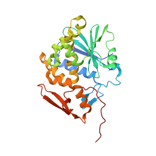
Conformational change in ricin toxin A-Chain: A critical factor for inhibitor binding to the secondary pocket.

Goto, M.Higashi, S.Ohba, T.Kawata, R.Nagatsu, K.Suzuki, S.Anslyn, E.V.Saito, R.

(2022) Biochem Biophys Res Commun 627: 1-4

  • DOI: https://doi.org/10.1016/j.bbrc.2022.08.008
  • Primary Citation of Related Structures:  
    7XZS, 7XZT, 7XZU, 7XZW, 7Y02, 7Y03, 7Y05, 7Y06, 7Y07, 7Y08

  • PubMed Abstract: 

    Ricin toxin A-chain (RTA), a toxic protein from Ricinus communis, inactivates ribosomes to induce toxicity. The active site of RTA consists of two binding pockets. Many studies have focused on developing RTA inhibitors that can simultaneously bind to these critical pockets; however, almost all the inhibitors developed so far interact with only one pocket. In the present study, we discovered that pterin-7-carboxamides with aromatic l-amino acid pendants interacted with the active site of the enzyme in a 2-to-1 mode, where one inhibitor molecule bound to the primary pocket and the second one entered the secondary pocket in the active site of RTA. X-ray crystallographic analysis of inhibitor/RTA complexes revealed that the conformational changes of Tyr80 and Asn122 in RTA were critical for triggering the entry of inhibitor molecules into the secondary pocket of the RTA active site.


  • Organizational Affiliation

    Department of Molecular Bioscience, Faculty of Science, Toho University, Funabashi, Chiba, 274-8510, Japan.


Macromolecules
Find similar proteins by:  (by identity cutoff)  |  3D Structure
Entity ID: 1
MoleculeChains Sequence LengthOrganismDetailsImage
Ricin A chain274Ricinus communisMutation(s): 0 
EC: 3.2.2.22
UniProt
Find proteins for P02879 (Ricinus communis)
Explore P02879 
Go to UniProtKB:  P02879
Entity Groups  
Sequence Clusters30% Identity50% Identity70% Identity90% Identity95% Identity100% Identity
UniProt GroupP02879
Sequence Annotations
Expand
  • Reference Sequence
Experimental Data & Validation

Experimental Data

  • Method: X-RAY DIFFRACTION
  • Resolution: 1.50 Å
  • R-Value Free: 0.243 
  • R-Value Work: 0.218 
  • R-Value Observed: 0.220 
  • Space Group: P 41 21 2
Unit Cell:
Length ( Å )Angle ( ˚ )
a = 67.46α = 90
b = 67.46β = 90
c = 141.06γ = 90
Software Package:
Software NamePurpose
XDSdata reduction
SCALAdata scaling
MOLREPphasing
REFMACrefinement
PDB_EXTRACTdata extraction

Structure Validation

View Full Validation Report


Ligand Structure Quality Assessment 


Entry History & Funding Information

Deposition Data


Funding OrganizationLocationGrant Number
Japan Society for the Promotion of Science (JSPS)JapanJP19K05699

Revision History  (Full details and data files)

  • Version 1.0: 2022-12-21
    Type: Initial release
  • Version 1.1: 2023-11-29
    Changes: Data collection, Refinement description