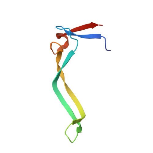
Structure of AcrVIA2 and its binding mechanism to CRISPR-Cas13a.
Song, G., Li, X., Wang, Z., Dong, C., Xie, X., Yan, X.(2022) Biochem Biophys Res Commun 612: 84-90
- PubMed: 35512461
- DOI: https://doi.org/10.1016/j.bbrc.2022.04.091
- Primary Citation of Related Structures:
7XMW - PubMed Abstract:
Phages and non-phage derived bacteria have evolved many anti-CRISPR proteins (Acrs) to escape the adaptive immune system of prokaryotes. Thus Acrs can be applied as a regulatory tool for gene edition by CRISPR system. Recently, a non-phage derived AcrVIA2 has been identified as an inhibitor that blocks the editing activity of Cas13a in vitro by binding to Cas13a. Here, we solved the crystal structure of AcrVIA2 at a resolution of 2.59 Å and confirmed that AcrVIA2 can bind to Helical-I domain in LshCas13a. Structural analysis show that the V-shaped acidic groove formed by β3-β3 hairpin of AcrVIA2 dimer is the key region that mediates the interaction between AcrVIA2 and Helical-I domain. In addition, we also reveal that Asp37 of AcrVIA2 plays an essential role in the functioning of the V-shaped acidic groove, and the functional dimer conformation of AcrVIA2 is stabilized by hydrogen bonds formed between Tyr41 of one monomer with Glu35 and Asp37 of the other monomer. These data expand the current understanding of the diverse interaction mechanisms between Acrs and Cas proteins, and also provide new ideas for the development of CRISPR-Cas13a regulatory tool.
- Department of Biochemistry and Molecular Biology, The Province and Ministry Co-sponsored Collaborative Innovation Center for Medical Epigenetics, Key Laboratory of Immune Microenvironment and Disease (Ministry of Education), School of Basic Medical Sciences, Tianjin Medical University, Tianjin, 300070, China.
Organizational Affiliation:


