7XJ7 | pdb_00007xj7

Crystal structure of engineered HIV-1 Reverse Transcriptase RNase H domain complexed with nitrofuran methoxy(methoxycarbonyl)phenyl ester


Experimental Data Snapshot

  • Method: X-RAY DIFFRACTION
  • Resolution: 1.80 Å
  • R-Value Free: 
    0.261 (Depositor), 0.257 (DCC) 
  • R-Value Work: 
    0.225 (Depositor), 0.225 (DCC) 
  • R-Value Observed: 
    0.227 (Depositor) 

Starting Model: experimental
View more details

wwPDB Validation 3D Report Full Report

Validation slider image for 7XJ7

This is version 1.3 of the entry. See complete history

Literature

Computational and Crystallographic Analysis of Binding Structures of Inhibitory Compounds for HIV-1 RNase H Activity.

Lu, H.Komukai, Y.Usami, K.Guo, Y.Qiao, X.Nukaga, M.Hoshino, T.

(2022) J Chem Inf Model 62: 6762-6774

  • DOI: https://doi.org/10.1021/acs.jcim.2c00537
  • Primary Citation Related Structures: 
    7XIS, 7XIT, 7XIU, 7XJ4, 7XJ5, 7XJ7

  • PubMed Abstract: 

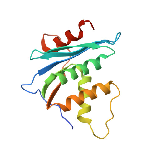
    Chemotherapy of human immunodeficiency virus type-1 (HIV-1) has significantly developed over the last three decades. The emergence of drug-resistant variants is, however, still a severe problem. The RNase H activity of HIV-1 reverse transcriptase is an attractive target for a new class of antiviral drugs because there is no approved inhibitor. The nitro-furan-carbonyl and nitro-thiophene-carbonyl groups are potent scaffolds for the HIV-1 RNase H inhibitor. In this work, the binding structures of six inhibitory compounds were obtained by X-ray crystal analysis in a complex with a recombinant protein of HIV-1 RNase H domain. Every inhibitory compound was found to be bound to the catalytic site with the furan- or thiophene-ring coordinated to two divalent metal ions at the binding pocket. All the atoms in nitro, furan, carbonyl, and two metals were aligned in the nitro-furan derivatives. The straight line connecting nitro and carboxyl groups was parallel to the plane made by two metal ions and a furan O atom. The binding modes of the nitro-thiophene derivatives were slightly different from those of the nitro-furan ones. The nitro and carbonyl groups deviated from the plane made by two metals and a thiophene S atom. Molecular dynamics simulations suggested that the furan O or thiophene S atom and carbonyl O atom were firmly coordinated to the metal ions. The simulations made the planar nitro-furan moiety well aligned to the line connecting the two metal ions. In contrast, the nitro-thiophene derivatives were displaced from the initial positions after the simulations. The computational findings will be a sound basis for developing potent inhibitors for HIV-1 RNase H activity.


  • Organizational Affiliation
    • Graduate School of Pharmaceutical Sciences, Chiba University Inohana 1-8-1, Chuo-ku, Chiba 260-8675, Japan.

Macromolecule Content 

  • Total Structure Weight: 17.27 kDa 
  • Atom Count: 1,224 
  • Modeled Residue Count: 145 
  • Deposited Residue Count: 151 
  • Unique protein chains: 1

Macromolecules

Find similar proteins by:|  3D Structure
Entity ID: 1
MoleculeChains  Sequence LengthOrganismDetailsImage
Reverse Transcriptase RNase H domain151Human immunodeficiency virus 1Mutation(s): 0 
EC: 3.1.26.13
UniProt
Find proteins for P04585 (Human immunodeficiency virus type 1 group M subtype B (isolate HXB2))
Explore P04585 
Go to UniProtKB:  P04585
Entity Groups
Sequence Clusters30% Identity50% Identity70% Identity90% Identity95% Identity100% Identity
UniProt GroupP04585
Sequence Annotations
Expand
Reference Sequence

Experimental Data & Validation

Experimental Data

  • Method: X-RAY DIFFRACTION
  • Resolution: 1.80 Å
  • R-Value Free:  0.261 (Depositor), 0.257 (DCC) 
  • R-Value Work:  0.225 (Depositor), 0.225 (DCC) 
  • R-Value Observed: 0.227 (Depositor) 
Space Group: P 41 21 2
Unit Cell:
Length ( Å )Angle ( ˚ )
a = 60.812α = 90
b = 60.812β = 90
c = 82.506γ = 90
Software Package:
Software NamePurpose
PHENIXrefinement
XDSdata reduction
Aimlessdata scaling
PDB_EXTRACTdata extraction
REFMACphasing

Structure Validation

View Full Validation Report


Entry History 

& Funding Information

Deposition Data


Funding OrganizationLocationGrant Number
Japan Society for the Promotion of Science (JSPS)Japan21K07050

Revision History  (Full details and data files)

  • Version 1.0: 2022-04-27
    Type: Initial release
  • Version 1.1: 2022-10-19
    Changes: Database references
  • Version 1.2: 2023-01-11
    Changes: Database references
  • Version 1.3: 2023-11-29
    Changes: Data collection, Refinement description