7XCL | pdb_00007xcl

Crystal structure of trimethylamine methyltransferase MttB from Methanosarcina barkeri at 2.5 A resolution


Experimental Data Snapshot

  • Method: X-RAY DIFFRACTION
  • Resolution: 2.50 Å
  • R-Value Free: 
    0.218 (Depositor), 0.195 (DCC) 
  • R-Value Work: 
    0.174 (Depositor), 0.193 (DCC) 

Starting Model: experimental
View more details

wwPDB Validation 3D Report Full Report

Validation slider image for 7XCL

This is version 1.2 of the entry. See complete history

Literature

Insights into pyrrolysine function from structures of a trimethylamine methyltransferase and its corrinoid protein complex.

Li, J.Kang, P.T.Jiang, R.Lee, J.Y.Soares, J.A.Krzycki, J.A.Chan, M.K.

(2023) Commun Biol 6: 54-54

  • DOI: https://doi.org/10.1038/s42003-022-04397-3
  • Primary Citation Related Structures: 
    7XCL, 7XCM, 7XCN

  • PubMed Abstract: 

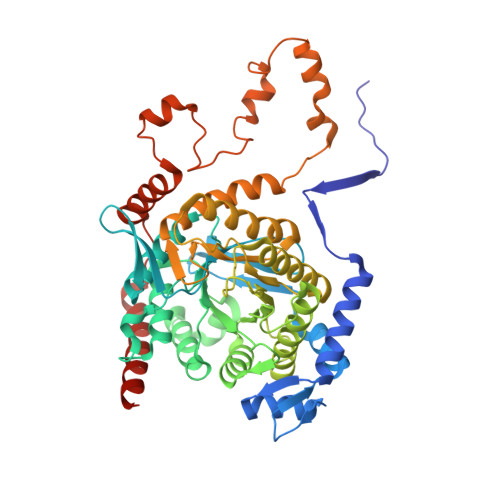
    The 22nd genetically encoded amino acid, pyrrolysine, plays a unique role in the key step in the growth of methanogens on mono-, di-, and tri-methylamines by activating the methyl group of these substrates for transfer to a corrinoid cofactor. Previous crystal structures of the Methanosarcina barkeri monomethylamine methyltransferase elucidated the structure of pyrrolysine and provide insight into its role in monomethylamine activation. Herein, we report the second structure of a pyrrolysine-containing protein, the M. barkeri trimethylamine methyltransferase MttB, and its structure bound to sulfite, a substrate analog of trimethylamine. We also report the structure of MttB in complex with its cognate corrinoid protein MttC, which specifically receives the methyl group from the pyrrolysine-activated trimethylamine substrate during methanogenesis. Together these structures provide key insights into the role of pyrrolysine in methyl group transfer from trimethylamine to the corrinoid cofactor in MttC.


  • Organizational Affiliation
    • School of Life Sciences, and Center of Novel Biomaterials, The Chinese University of Hong Kong, Shatin, Hong Kong, China.

Macromolecule Content 

  • Total Structure Weight: 330.28 kDa 
  • Atom Count: 23,762 
  • Modeled Residue Count: 2,963 
  • Deposited Residue Count: 3,018 
  • Unique protein chains: 1

Macromolecules

Find similar proteins by:|  3D Structure
Entity ID: 1
MoleculeChains  Sequence LengthOrganismDetailsImage
Trimethylamine methyltransferase
A, B, C, D, E
A, B, C, D, E, F
503Methanosarcina barkeri MSMutation(s): 0 
Gene Names: MSBRM_0461
EC: 2.1.1.250
UniProt
Find proteins for O93658 (Methanosarcina barkeri)
Explore O93658 
Go to UniProtKB:  O93658
Entity Groups
Sequence Clusters30% Identity50% Identity70% Identity90% Identity95% Identity100% Identity
UniProt GroupO93658
Sequence Annotations
Expand
Reference Sequence

Small Molecules

Ligands 2 Unique
IDChains Name / Formula / InChI Key2D Diagram3D Interactions
GOL

Query on GOL


Download:Ideal Coordinates CCD File
G [auth A]
H [auth A]
J [auth B]
K [auth B]
M [auth C]
G [auth A],
H [auth A],
J [auth B],
K [auth B],
M [auth C],
O [auth D],
P [auth D],
Q [auth D],
S [auth E],
T [auth E],
V [auth F]
GLYCEROL
C3 H8 O3
PEDCQBHIVMGVHV-UHFFFAOYSA-N
NA

Query on NA


Download:Ideal Coordinates CCD File
I [auth A]
L [auth B]
N [auth C]
R [auth D]
U [auth E]
I [auth A],
L [auth B],
N [auth C],
R [auth D],
U [auth E],
W [auth F]
SODIUM ION
Na
FKNQFGJONOIPTF-UHFFFAOYSA-N

Experimental Data & Validation

Experimental Data

  • Method: X-RAY DIFFRACTION
  • Resolution: 2.50 Å
  • R-Value Free:  0.218 (Depositor), 0.195 (DCC) 
  • R-Value Work:  0.174 (Depositor), 0.193 (DCC) 
Space Group: P 32 2 1
Unit Cell:
Length ( Å )Angle ( ˚ )
a = 175.492α = 90
b = 175.492β = 90
c = 300.996γ = 120
Software Package:
Software NamePurpose
CNSrefinement
HKL-2000data scaling
PDB_EXTRACTdata extraction
HKL-2000data reduction
PHASERphasing

Structure Validation

View Full Validation Report


Entry History 

& Funding Information

Deposition Data


Funding OrganizationLocationGrant Number
The University Grants Committee, Research Grants Council (RGC)Hong Kong14116620

Revision History  (Full details and data files)

  • Version 1.0: 2023-01-18
    Type: Initial release
  • Version 1.1: 2023-02-01
    Changes: Database references
  • Version 1.2: 2023-11-29
    Changes: Data collection, Refinement description