


Structural insights of 4-Hydrophenylpyruvate dioxygenase inhibition by structurally diverse small molecules
Dong, J., Dong, J., Yu, X.H., Yan, Y.C., Nan, J.X., He, B., Ye, B.Q., Yang, W.C., Lin, H.Y., Yang, G.F.(2022) Adv Agrochem
Experimental Data Snapshot
Starting Model: experimental
View more details
(2022) Adv Agrochem
Entity ID: 1 | |||||
|---|---|---|---|---|---|
| Molecule | Chains | Sequence Length | Organism | Details | Image |
| 4-hydroxyphenylpyruvate dioxygenase | 444 | Zea mays | Mutation(s): 1 Gene Names: ZEAMMB73_Zm00001d015356 | 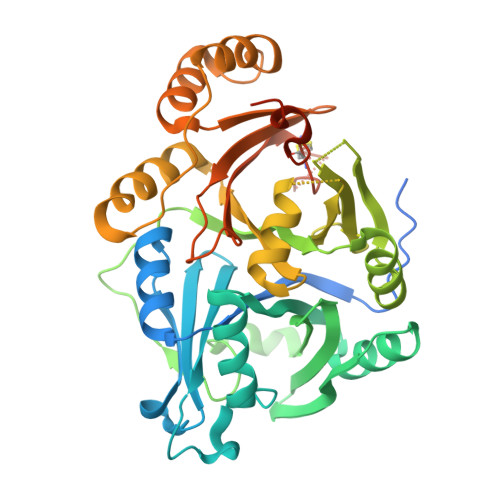 | |
UniProt | |||||
Entity Groups | |||||
| Sequence Clusters | 30% Identity50% Identity70% Identity90% Identity95% Identity100% Identity | ||||
| UniProt Group | A0A1D6H1G0 | ||||
Sequence AnnotationsExpand | |||||
Reference Sequence | |||||
| Ligands 2 Unique | |||||
|---|---|---|---|---|---|
| ID | Chains | Name / Formula / InChI Key | 2D Diagram | 3D Interactions | |
| 94L Download:Ideal Coordinates CCD File | F [auth A], H [auth B], J [auth C], L [auth D] | 3-(2,6-dimethylphenyl)-1-methyl-6-(2-oxidanyl-6-oxidanylidene-cyclohexen-1-yl)carbonyl-quinazoline-2,4-dione C24 H22 N2 O5 IFHRYUOSJUERAZ-UHFFFAOYSA-N |  | ||
| CO Download:Ideal Coordinates CCD File | E [auth A], G [auth B], I [auth C], K [auth D] | COBALT (II) ION Co XLJKHNWPARRRJB-UHFFFAOYSA-N |  | ||
| Length ( Å ) | Angle ( ˚ ) |
|---|---|
| a = 53.952 | α = 89.78 |
| b = 83.428 | β = 97.46 |
| c = 85.61 | γ = 107.64 |
| Software Name | Purpose |
|---|---|
| PHENIX | refinement |
| XDS | data reduction |
| XDS | data scaling |
| PHASER | phasing |
| Funding Organization | Location | Grant Number |
|---|---|---|
| National Science Foundation (NSF, China) | China | -- |


