


Crystal structure of full-length peptidyl-tRNA hydrolase from Mycobacterium tuberculosis
Kulandaisamy, R., Das, U., Inampudi, K.K.To be published.
Experimental Data Snapshot
Starting Model: experimental
View more details
wwPDB Validation 3D Report Full Report

Entity ID: 1 | |||||
|---|---|---|---|---|---|
| Molecule | Chains | Sequence Length | Organism | Details | Image |
| Peptidyl-tRNA hydrolase | 202 | Mycobacteriaceae bacterium | Mutation(s): 0 Gene Names: pth EC: 3.1.1.29 | 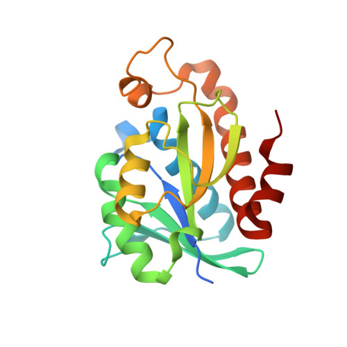 | |
UniProt | |||||
Entity Groups | |||||
| Sequence Clusters | 30% Identity50% Identity70% Identity90% Identity95% Identity100% Identity | ||||
| UniProt Group | P9WHN7 | ||||
Sequence AnnotationsExpand | |||||
Reference Sequence | |||||
| Length ( Å ) | Angle ( ˚ ) |
|---|---|
| a = 36.581 | α = 90 |
| b = 39.003 | β = 90 |
| c = 123.818 | γ = 90 |
| Software Name | Purpose |
|---|---|
| PHENIX | refinement |
| XDS | data reduction |
| XSCALE | data scaling |
| REFMAC | phasing |
| Funding Organization | Location | Grant Number |
|---|---|---|
| Department of Science & Technology (DST, India) | India | EEQ/2016/000507 |
| Department of Biotechnology (DBT, India) | India | BT/PR34319/Med/29/1488/2019 |


