7WSZ

human glyoxalase I (with C-ter His tag) in glycerol-bound form


Experimental Data Snapshot

  • Method: X-RAY DIFFRACTION
  • Resolution: 1.52 Å
  • R-Value Free: 0.187 
  • R-Value Work: 0.161 
  • R-Value Observed: 0.164 

wwPDB Validation   3D Report Full Report


This is version 1.2 of the entry. See complete history


Literature

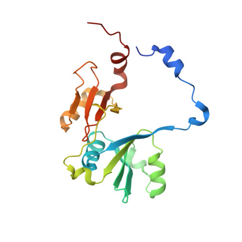
Crystal structures of human glyoxalase I and its complex with TLSC702 reveal inhibitor binding mode and substrate preference.

Usami, M.Ando, K.Shibuya, A.Takasawa, R.Yokoyama, H.

(2022) FEBS Lett 596: 1458-1467

  • DOI: https://doi.org/10.1002/1873-3468.14344
  • Primary Citation of Related Structures:  
    7WSZ, 7WT0, 7WT1, 7WT2

  • PubMed Abstract: 

    Human glyoxalase I (hGLO I) is an enzyme for detoxification of methylglyoxal (MG) and has been considered an attractive target for the development of new anticancer drugs. In our previous report, the GLO I inhibitor TLSC702 induced apoptosis in tumor cells. Here, we determined the crystal structures of hGLO I and its complex with TLSC702. In the complex, the carboxyl O atom of TLSC702 is coordinated to Zn 2+ , and TLSC702 mainly shows van der Waals interaction with hydrophobic residues. In the inhibitor-unbound structure, glycerol, which has similar functional groups to MG, was bound to Zn 2+ , indicating that GLO I can easily bind to MG. This study provides a structural basis to develop better anticancer drugs.


  • Organizational Affiliation

    Faculty of Pharmaceutical Sciences, Tokyo University of Science, Noda, Japan.


Macromolecules
Find similar proteins by:  (by identity cutoff)  |  3D Structure
Entity ID: 1
MoleculeChains Sequence LengthOrganismDetailsImage
Lactoylglutathione lyase
A, B
192Homo sapiensMutation(s): 0 
Gene Names: GLO1
EC: 4.4.1.5
UniProt & NIH Common Fund Data Resources
Find proteins for Q04760 (Homo sapiens)
Explore Q04760 
Go to UniProtKB:  Q04760
PHAROS:  Q04760
GTEx:  ENSG00000124767 
Entity Groups  
Sequence Clusters30% Identity50% Identity70% Identity90% Identity95% Identity100% Identity
UniProt GroupQ04760
Sequence Annotations
Expand
  • Reference Sequence
Small Molecules
Ligands 5 Unique
IDChains Name / Formula / InChI Key2D Diagram3D Interactions
GOL
Query on GOL

Download Ideal Coordinates CCD File 
D [auth A],
E [auth A],
F [auth A],
M [auth B]
GLYCEROL
C3 H8 O3
PEDCQBHIVMGVHV-UHFFFAOYSA-N
BME
Query on BME

Download Ideal Coordinates CCD File 
J [auth A],
K [auth A]
BETA-MERCAPTOETHANOL
C2 H6 O S
DGVVWUTYPXICAM-UHFFFAOYSA-N
IMD
Query on IMD

Download Ideal Coordinates CCD File 
I [auth A]IMIDAZOLE
C3 H5 N2
RAXXELZNTBOGNW-UHFFFAOYSA-O
ZN
Query on ZN

Download Ideal Coordinates CCD File 
C [auth A],
L [auth B]
ZINC ION
Zn
PTFCDOFLOPIGGS-UHFFFAOYSA-N
BO3
Query on BO3

Download Ideal Coordinates CCD File 
G [auth A],
H [auth A],
N [auth B]
BORIC ACID
B H3 O3
KGBXLFKZBHKPEV-UHFFFAOYSA-N
Experimental Data & Validation

Experimental Data

  • Method: X-RAY DIFFRACTION
  • Resolution: 1.52 Å
  • R-Value Free: 0.187 
  • R-Value Work: 0.161 
  • R-Value Observed: 0.164 
  • Space Group: P 21 21 21
Unit Cell:
Length ( Å )Angle ( ˚ )
a = 85.225α = 90
b = 96.995β = 90
c = 46.264γ = 90
Software Package:
Software NamePurpose
REFMACrefinement
PDB_EXTRACTdata extraction
XDSdata reduction
SCALAdata scaling
MOLREPphasing

Structure Validation

View Full Validation Report


Entry History & Funding Information

Deposition Data


Funding OrganizationLocationGrant Number
Japan Society for the Promotion of Science (JSPS)Japan17K07316

Revision History  (Full details and data files)

  • Version 1.0: 2022-04-13
    Type: Initial release
  • Version 1.1: 2022-06-29
    Changes: Database references
  • Version 1.2: 2023-11-29
    Changes: Data collection, Refinement description