7WEK

Crystal structure of the mouse Wdr47 NTD in complex with the WBR motif form Camsap3.


Experimental Data Snapshot

  • Method: X-RAY DIFFRACTION
  • Resolution: 3.21 Å
  • R-Value Free: 0.314 
  • R-Value Work: 0.271 
  • R-Value Observed: 0.275 

wwPDB Validation   3D Report Full Report


This is version 1.1 of the entry. See complete history


Literature

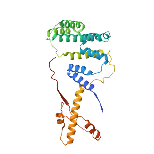
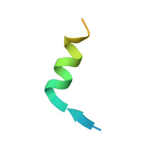
Intertwined Wdr47-NTD dimer recognizes a basic-helical motif in Camsap proteins for proper central-pair microtubule formation.

Ren, J.Li, D.Liu, J.Liu, H.Yan, X.Zhu, X.Feng, W.

(2022) Cell Rep 41: 111589-111589

  • DOI: https://doi.org/10.1016/j.celrep.2022.111589
  • Primary Citation of Related Structures:  
    7WEJ, 7WEK

  • PubMed Abstract: 

    Calmodulin-regulated spectrin-associated proteins (Camsaps) bind to the N-terminal domain of WD40-repeat 47 (Wdr47-NTD; featured with a LisH-CTLH motif) to properly generate axonemal central-pair microtubules (CP-MTs) for the planar beat pattern of mammalian motile multicilia. The underlying molecular mechanism, however, remains unclear. Here, we determine the structures of apo-Wdr47-NTD and Wdr47-NTD in complex with a characteristic Wdr47-binding region (WBR) from Camsap3. Wdr47-NTD forms an intertwined dimer with a special cross-over region (COR) in addition to the canonical LisH and globular α-helical core (GAC). The basic WBR peptide adopts an α-helical conformation and anchors to a tailored acidic pocket embedded in the COR. Mutations in this target-binding pocket disrupt the interaction between Wdr47-NTD and Camsap3. Impairing Wdr47-Camsap interactions markedly reduces rescue effects of Wdr47 on CP-MTs and ciliary beat of Wdr47-deficient ependymal cells. Thus, Wdr47-NTD functions by recognizing a specific basic helical motif in Camsap proteins via its non-canonical COR, a target-binding site in LisH-CTLH-containing domains.


  • Organizational Affiliation

    National Laboratory of Biomacromolecules, CAS Center for Excellence in Biomacromolecules, Institute of Biophysics, Chinese Academy of Sciences, 15 Datun Road, Beijing 100101, China.


Macromolecules
Find similar proteins by:  (by identity cutoff)  |  3D Structure
Entity ID: 1
MoleculeChains Sequence LengthOrganismDetailsImage
WD repeat-containing protein 47
A, B
295Mus musculusMutation(s): 0 
Gene Names: Wdr47Kiaa0893
UniProt & NIH Common Fund Data Resources
Find proteins for Q8CGF6 (Mus musculus)
Explore Q8CGF6 
Go to UniProtKB:  Q8CGF6
IMPC:  MGI:2139593
Entity Groups  
Sequence Clusters30% Identity50% Identity70% Identity90% Identity95% Identity100% Identity
UniProt GroupQ8CGF6
Sequence Annotations
Expand
  • Reference Sequence
Find similar proteins by:  (by identity cutoff)  |  3D Structure
Entity ID: 2
MoleculeChains Sequence LengthOrganismDetailsImage
WBR motif form Calmodulin-regulated spectrin-associated protein 3
C, D
30Mus musculusMutation(s): 0 
UniProt & NIH Common Fund Data Resources
Find proteins for Q80VC9 (Mus musculus)
Explore Q80VC9 
Go to UniProtKB:  Q80VC9
IMPC:  MGI:1916947
Entity Groups  
Sequence Clusters30% Identity50% Identity70% Identity90% Identity95% Identity100% Identity
UniProt GroupQ80VC9
Sequence Annotations
Expand
  • Reference Sequence
Experimental Data & Validation

Experimental Data

  • Method: X-RAY DIFFRACTION
  • Resolution: 3.21 Å
  • R-Value Free: 0.314 
  • R-Value Work: 0.271 
  • R-Value Observed: 0.275 
  • Space Group: P 21 21 21
Unit Cell:
Length ( Å )Angle ( ˚ )
a = 58.077α = 90
b = 105.318β = 90
c = 155.972γ = 90
Software Package:
Software NamePurpose
PHENIXrefinement
PDB_EXTRACTdata extraction
HKL-2000data reduction
HKL-2000data scaling
PHASERphasing

Structure Validation

View Full Validation Report


Entry History & Funding Information

Deposition Data


Funding OrganizationLocationGrant Number
National Science Foundation (NSF, China)China--

Revision History  (Full details and data files)

  • Version 1.0: 2022-11-30
    Type: Initial release
  • Version 1.1: 2023-11-29
    Changes: Data collection, Refinement description