7WAD

Trichodesmium erythraeum cyanophycin synthetase 1 (TeCphA1) with ATPgammaS


Experimental Data Snapshot

  • Method: ELECTRON MICROSCOPY
  • Resolution: 2.96 Å
  • Aggregation State: PARTICLE 
  • Reconstruction Method: SINGLE PARTICLE 

wwPDB Validation   3D Report Full Report


This is version 1.1 of the entry. See complete history


Literature

Structural bases for aspartate recognition and polymerization efficiency of cyanobacterial cyanophycin synthetase.

Miyakawa, T.Yang, J.Kawasaki, M.Adachi, N.Fujii, A.Miyauchi, Y.Muramatsu, T.Moriya, T.Senda, T.Tanokura, M.

(2022) Nat Commun 13: 5097-5097

  • DOI: https://doi.org/10.1038/s41467-022-32834-8
  • Primary Citation of Related Structures:  
    7WAC, 7WAD, 7WAE, 7WAF

  • PubMed Abstract: 

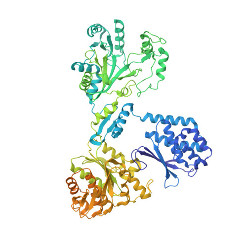
    Cyanophycin is a natural biopolymer consisting of equimolar amounts of aspartate and arginine as the backbone and branched sidechain, respectively. It is produced by a single enzyme, cyanophycin synthetase (CphA1), and accumulates as a nitrogen reservoir during N 2 fixation by most cyanobacteria. A recent structural study showed that three constituent domains of CphA1 function as two distinct catalytic sites and an oligomerization interface in cyanophycin synthesis. However, it remains unclear how the ATP-dependent addition of aspartate to cyanophycin is initiated at the catalytic site of the glutathione synthetase-like domain. Here, we report the cryogenic electron microscopy structures of CphA1, including a complex with aspartate, cyanophycin primer peptide, and ATP analog. These structures reveal the aspartate binding mode and phosphate-binding loop movement to the active site required for the reaction. Furthermore, structural and mutational data show a potential role of protein dynamics in the catalytic efficiency of the arginine condensation reaction.


  • Organizational Affiliation

    Department of Applied Biological Chemistry, Graduate School of Agricultural and Life Sciences, The University of Tokyo, 1-1-1 Yayoi, Bunkyo-ku, Tokyo, 113-8657, Japan.


Macromolecules
Find similar proteins by:  (by identity cutoff)  |  3D Structure
Entity ID: 1
MoleculeChains Sequence LengthOrganismDetailsImage
Cyanophycin synthase
A, B, C, D
910Trichodesmium erythraeum IMS101Mutation(s): 0 
Gene Names: Tery_1965
EC: 6.3.2.29 (PDB Primary Data), 6.3.2.30 (PDB Primary Data)
UniProt
Find proteins for Q113V7 (Trichodesmium erythraeum (strain IMS101))
Explore Q113V7 
Go to UniProtKB:  Q113V7
Entity Groups  
Sequence Clusters30% Identity50% Identity70% Identity90% Identity95% Identity100% Identity
UniProt GroupQ113V7
Sequence Annotations
Expand
  • Reference Sequence
Small Molecules
Ligands 2 Unique
IDChains Name / Formula / InChI Key2D Diagram3D Interactions
AGS (Subject of Investigation/LOI)
Query on AGS

Download Ideal Coordinates CCD File 
F [auth A]
G [auth A]
I [auth B]
J [auth B]
L [auth C]
F [auth A],
G [auth A],
I [auth B],
J [auth B],
L [auth C],
M [auth C],
O [auth D],
P [auth D]
PHOSPHOTHIOPHOSPHORIC ACID-ADENYLATE ESTER
C10 H16 N5 O12 P3 S
NLTUCYMLOPLUHL-KQYNXXCUSA-N
MG (Subject of Investigation/LOI)
Query on MG

Download Ideal Coordinates CCD File 
E [auth A],
H [auth B],
K [auth C],
N [auth D]
MAGNESIUM ION
Mg
JLVVSXFLKOJNIY-UHFFFAOYSA-N
Experimental Data & Validation

Experimental Data

  • Method: ELECTRON MICROSCOPY
  • Resolution: 2.96 Å
  • Aggregation State: PARTICLE 
  • Reconstruction Method: SINGLE PARTICLE 
EM Software:
TaskSoftware PackageVersion
MODEL REFINEMENTCoot0.8.9.2
MODEL REFINEMENTPHENIX1.19.2
RECONSTRUCTIONRELION3.1

Structure Validation

View Full Validation Report


Entry History & Funding Information

Deposition Data


Funding OrganizationLocationGrant Number
Japan Agency for Medical Research and Development (AMED)JapanJP21am0101077
Japan Agency for Medical Research and Development (AMED)JapanJP21am0101071

Revision History  (Full details and data files)

  • Version 1.0: 2022-09-07
    Type: Initial release
  • Version 1.1: 2022-09-14
    Changes: Database references