7W7A

Heme exporter in complex with Mn-containing protoporphyrin IX, Mn-anomalous data


Experimental Data Snapshot

  • Method: X-RAY DIFFRACTION
  • Resolution: 3.20 Å
  • R-Value Free: 0.303 
  • R-Value Work: 0.264 
  • R-Value Observed: 0.265 

wwPDB Validation   3D Report Full Report


Ligand Structure Quality Assessment 


This is version 1.2 of the entry. See complete history


Literature

Structural basis for heme detoxification by an ATP-binding cassette-type efflux pump in gram-positive pathogenic bacteria.

Nakamura, H.Hisano, T.Rahman, M.M.Tosha, T.Shirouzu, M.Shiro, Y.

(2022) Proc Natl Acad Sci U S A 119: e2123385119-e2123385119

  • DOI: https://doi.org/10.1073/pnas.2123385119
  • Primary Citation of Related Structures:  
    7W78, 7W79, 7W7A, 7W7B, 7W7C, 7W7D

  • PubMed Abstract: 

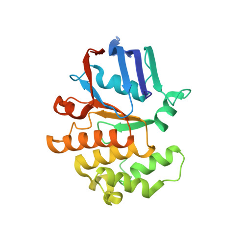
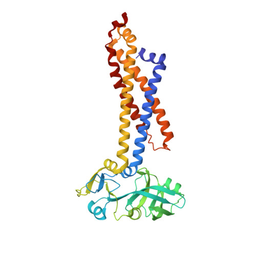
    Bacterial pathogens acquire heme from the host hemoglobin as an iron nutrient for their virulence and proliferation in blood. Concurrently, they encounter cytotoxic-free heme that escapes the heme-acquisition process. To overcome this toxicity, many gram-positive bacteria employ an ATP-binding cassette heme-dedicated efflux pump, HrtBA in the cytoplasmic membranes. Although genetic analyses have suggested that HrtBA expels heme from the bacterial membranes, the molecular mechanism of heme efflux remains elusive due to the lack of protein studies. Here, we show the biochemical properties and crystal structures of Corynebacterium diphtheriae HrtBA, alone and in complex with heme or an ATP analog, and we reveal how HrtBA extracts heme from the membrane and releases it. HrtBA consists of two cytoplasmic HrtA ATPase subunits and two transmembrane HrtB permease subunits. A heme-binding site is formed in the HrtB dimer and is laterally accessible to heme in the outer leaflet of the membrane. The heme-binding site captures heme from the membrane using a glutamate residue of either subunit as an axial ligand and sequesters the heme within the rearranged transmembrane helix bundle. By ATP-driven HrtA dimerization, the heme-binding site is squeezed to extrude the bound heme. The mechanism sheds light on the detoxification of membrane-bound heme in this bacterium.


  • Organizational Affiliation

    Laboratory for Protein Functional and Structural Biology, RIKEN Center for Biosystems Dynamics Research, Yokohama 230-0045, Japan.


Macromolecules
Find similar proteins by:  (by identity cutoff)  |  3D Structure
Entity ID: 1
MoleculeChains Sequence LengthOrganismDetailsImage
Putative ABC transport system, ATP-binding protein
A, C, E, G, I
A, C, E, G, I, K
231Corynebacterium diphtheriae NCTC 13129Mutation(s): 0 
Gene Names: DIP2323
UniProt
Find proteins for Q6NEF2 (Corynebacterium diphtheriae (strain ATCC 700971 / NCTC 13129 / Biotype gravis))
Explore Q6NEF2 
Go to UniProtKB:  Q6NEF2
Entity Groups  
Sequence Clusters30% Identity50% Identity70% Identity90% Identity95% Identity100% Identity
UniProt GroupQ6NEF2
Sequence Annotations
Expand
  • Reference Sequence
Find similar proteins by:  (by identity cutoff)  |  3D Structure
Entity ID: 2
MoleculeChains Sequence LengthOrganismDetailsImage
Putative ABC transport system integral membrane protein
B, D, F, H, J
B, D, F, H, J, L
344Corynebacterium diphtheriae NCTC 13129Mutation(s): 0 
Gene Names: DIP2324
Membrane Entity: Yes 
UniProt
Find proteins for Q6NEF1 (Corynebacterium diphtheriae (strain ATCC 700971 / NCTC 13129 / Biotype gravis))
Explore Q6NEF1 
Go to UniProtKB:  Q6NEF1
Entity Groups  
Sequence Clusters30% Identity50% Identity70% Identity90% Identity95% Identity100% Identity
UniProt GroupQ6NEF1
Sequence Annotations
Expand
  • Reference Sequence
Small Molecules
Ligands 5 Unique
IDChains Name / Formula / InChI Key2D Diagram3D Interactions
MNR (Subject of Investigation/LOI)
Query on MNR

Download Ideal Coordinates CCD File 
O [auth D],
Q [auth H]
PROTOPORPHYRIN IX CONTAINING MN
C34 H32 Mn N4 O4
UAPHVASSHGRKEN-RGGAHWMASA-L
AGA
Query on AGA

Download Ideal Coordinates CCD File 
M [auth B](1S)-2-{[{[(2S)-2,3-DIHYDROXYPROPYL]OXY}(HYDROXY)PHOSPHORYL]OXY}-1-[(PENTANOYLOXY)METHYL]ETHYL OCTANOATE
C19 H36 O10 P
UQSXQYRZHMGKIE-DLBZAZTESA-M
PGE
Query on PGE

Download Ideal Coordinates CCD File 
N [auth B]TRIETHYLENE GLYCOL
C6 H14 O4
ZIBGPFATKBEMQZ-UHFFFAOYSA-N
PEG
Query on PEG

Download Ideal Coordinates CCD File 
P [auth D]DI(HYDROXYETHYL)ETHER
C4 H10 O3
MTHSVFCYNBDYFN-UHFFFAOYSA-N
EDO
Query on EDO

Download Ideal Coordinates CCD File 
R [auth H]1,2-ETHANEDIOL
C2 H6 O2
LYCAIKOWRPUZTN-UHFFFAOYSA-N
Experimental Data & Validation

Experimental Data

  • Method: X-RAY DIFFRACTION
  • Resolution: 3.20 Å
  • R-Value Free: 0.303 
  • R-Value Work: 0.264 
  • R-Value Observed: 0.265 
  • Space Group: P 1
Unit Cell:
Length ( Å )Angle ( ˚ )
a = 82.488α = 111.81
b = 133.672β = 99.83
c = 159.838γ = 94.43
Software Package:
Software NamePurpose
XSCALEdata scaling
PHENIXrefinement
PDB_EXTRACTdata extraction
XDSdata reduction
PHENIXphasing

Structure Validation

View Full Validation Report


Ligand Structure Quality Assessment 


Entry History & Funding Information

Deposition Data


Funding OrganizationLocationGrant Number
Japan Society for the Promotion of Science (JSPS)Japan26220807
Japan Society for the Promotion of Science (JSPS)Japan16K07309
Japan Society for the Promotion of Science (JSPS)JapanJP19H00926
Japan Society for the Promotion of Science (JSPS)JapanJP19H05761

Revision History  (Full details and data files)

  • Version 1.0: 2022-06-22
    Type: Initial release
  • Version 1.1: 2022-12-21
    Changes: Database references
  • Version 1.2: 2023-11-29
    Changes: Data collection, Refinement description