Identification, structure determination and analysis of Mycobacterium smegmatis acyl-carrier protein synthase (AcpS) crystallized serendipitously.
Bhatia, I., Yadav, S., Biswal, B.K.(2022) Acta Crystallogr F Struct Biol Commun 78: 252-264
- PubMed: 35787552 Search on PubMedSearch on PubMed Central
- DOI: https://doi.org/10.1107/S2053230X22005738
- Primary Citation Related Structures:
7W58 - PubMed Abstract:
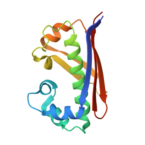
The unintended crystallization of proteins which generally originate from the expression host instead of the target recombinant proteins is periodically reported. Despite the massive technological advances in the field, assigning a structural model to the corresponding diffraction data is not a trivial task. Here, the structure of acyl-carrier protein synthase (AcpS) from Mycobacterium smegmatis (msAcpS), which crystallized inadvertently in an experimental setup to grow crystals of a Mycobacterium tuberculosis protein using M. smegmatis as an expression system, is reported. After numerous unsuccessful attempts to solve the structure of the target protein by the molecular-replacement method no convincing solutions were obtained, indicating that the diffraction data may correspond to a crystal of an artifactual protein, which was finally identified by the Sequence-Independent Molecular replacement Based on Available Databases (SIMBAD) server. The msAcpS structure was solved at 2.27 Å resolution and structural analysis showed an overall conserved fold. msAcpS formed a trimeric structure similar to those of other reported structures of AcpS from various organisms; however, the residues involved in trimer formation are not strictly conserved. An unrelated metal ion (Ni 2+ ), which was possibly incorporated during protein purification, was observed in the proximity of His49 and His116. Structural and sequence differences were observed in the loop connecting the α3 and α4 helices that is responsible for the open and closed conformations of the enzyme. Moreover, the structural analysis of msAcpS augments the current understanding of this enzyme, which plays a crucial role in the functional activation of acyl-carrier proteins in the fatty-acid biosynthesis pathway.
- Structural and Functional Biology Laboratory, National Institute of Immunology, Aruna Asaf Ali Marg, New Delhi 110 067, India.
Organizational Affiliation: