7W54

Crystal structure of a bacterial OTU DUB with Ub-PA


Experimental Data Snapshot

  • Method: X-RAY DIFFRACTION
  • Resolution: 2.64 Å
  • R-Value Free: 0.253 
  • R-Value Work: 0.216 
  • R-Value Observed: 0.218 

wwPDB Validation   3D Report Full Report


Ligand Structure Quality Assessment 


This is version 1.2 of the entry. See complete history


Literature

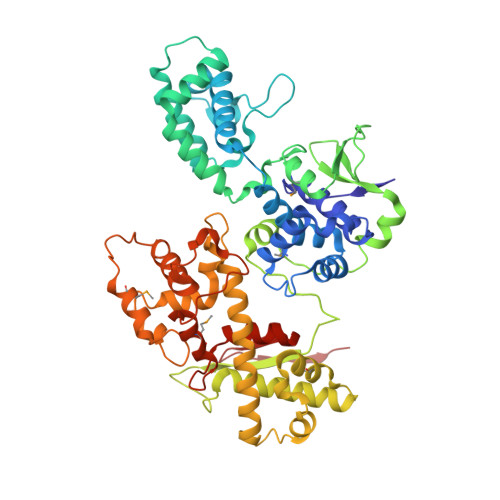
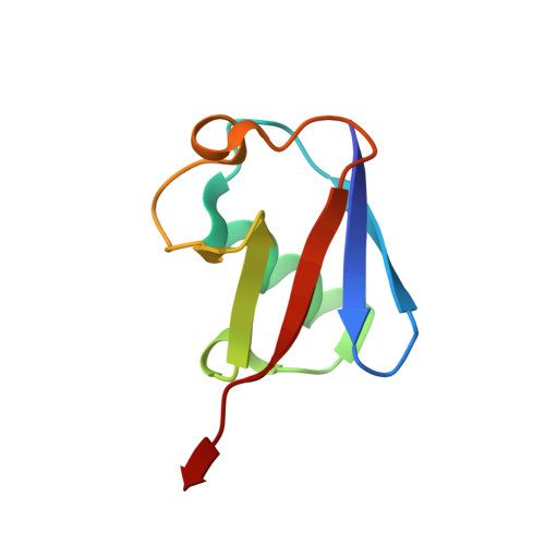
Structural basis for the dual catalytic activity of the Legionella pneumophila ovarian tumor (OTU) domain deubiquitinase LotA.

Luo, J.Ruan, X.Huang, Z.Li, Z.Ye, L.Wu, Y.Zhen, X.Ouyang, S.

(2022) J Biol Chem 298: 102414-102414

  • DOI: https://doi.org/10.1016/j.jbc.2022.102414
  • Primary Citation of Related Structures:  
    7W54

  • PubMed Abstract: 

    Legionella pneumophila, a bacterial pathogen that causes a severe pneumonia known as Legionnaires' disease, extensively exploits the ubiquitin (Ub) pathway in the infected host cells through certain virulence effectors excreted by the Dot/Icm system. To date, several Dot/Icm effectors have been found to act as Ub ligases, and four effectors, including LotA, LotB, LotC, and Ceg7, have been identified as deubiquitinases (DUBs) from the ovarian tumor (OTU) domain family. LotA is unique among other OTU DUBs because it possesses two distinct DUB domains and exclusively exhibits catalytic activity against K6-linked diUb and polyUb chains. However, the structure of LotA and the molecular mechanism for the dual DUB activity remains elusive. In this study, we solved the structure of LotA in complex with proximally bound Ub and distal covalently bound Ub. Both Ub molecules are bound to the DUB1 domain and mimic a K6-linked diUb. Structural analysis reveals that the DUB1 domain utilizes a distinct mechanism for recognition of the K6-linked diUb within a large S1' binding site that is uncommon to OTU DUBs. Structural fold of the LotA DUB2 domain closely resembles LotB and LotC, similarly containing an extra α-helix lobe that has been demonstrated to play an important role in Ub binding. Collectively, our study uncovers the structural basis for the dual catalytic activity of the unique OTU family DUB LotA.


  • Organizational Affiliation

    Provincial University Key Laboratory of Cellular Stress Response and Metabolic Regulation, The Key Laboratory of Innate Immune Biology of Fujian Province, Biomedical Research Center of South China, Key Laboratory of OptoElectronic Science and Technology for Medicine of the Ministry of Education, College of Life Sciences, Fujian Normal University, Fuzhou, China.


Macromolecules
Find similar proteins by:  (by identity cutoff)  |  3D Structure
Entity ID: 1
MoleculeChains Sequence LengthOrganismDetailsImage
Lpg2248
A, B
521Legionella pneumophila subsp. pneumophila str. Philadelphia 1Mutation(s): 0 
Gene Names: lpg2248
UniProt
Find proteins for Q5ZTB4 (Legionella pneumophila subsp. pneumophila (strain Philadelphia 1 / ATCC 33152 / DSM 7513))
Explore Q5ZTB4 
Go to UniProtKB:  Q5ZTB4
Entity Groups  
Sequence Clusters30% Identity50% Identity70% Identity90% Identity95% Identity100% Identity
UniProt GroupQ5ZTB4
Sequence Annotations
Expand
  • Reference Sequence
Find similar proteins by:  (by identity cutoff)  |  3D Structure
Entity ID: 2
MoleculeChains Sequence LengthOrganismDetailsImage
Polyubiquitin-B
C, D, E, F
75Homo sapiensMutation(s): 0 
Gene Names: UBB
UniProt & NIH Common Fund Data Resources
Find proteins for P0CG47 (Homo sapiens)
Explore P0CG47 
Go to UniProtKB:  P0CG47
PHAROS:  P0CG47
GTEx:  ENSG00000170315 
Entity Groups  
Sequence Clusters30% Identity50% Identity70% Identity90% Identity95% Identity100% Identity
UniProt GroupP0CG47
Sequence Annotations
Expand
  • Reference Sequence
Small Molecules
Modified Residues  1 Unique
IDChains TypeFormula2D DiagramParent
MSE
Query on MSE
A, B
L-PEPTIDE LINKINGC5 H11 N O2 SeMET
Experimental Data & Validation

Experimental Data

  • Method: X-RAY DIFFRACTION
  • Resolution: 2.64 Å
  • R-Value Free: 0.253 
  • R-Value Work: 0.216 
  • R-Value Observed: 0.218 
  • Space Group: P 21 21 21
Unit Cell:
Length ( Å )Angle ( ˚ )
a = 73.65α = 90
b = 126.09β = 90
c = 207.09γ = 90
Software Package:
Software NamePurpose
REFMACrefinement
HKL-2000data scaling
PDB_EXTRACTdata extraction
PHENIXmodel building
AutoSolphasing

Structure Validation

View Full Validation Report


Ligand Structure Quality Assessment 


Entry History & Funding Information

Deposition Data


Funding OrganizationLocationGrant Number
National Natural Science Foundation of China (NSFC)China--

Revision History  (Full details and data files)

  • Version 1.0: 2022-08-24
    Type: Initial release
  • Version 1.1: 2023-01-11
    Changes: Database references
  • Version 1.2: 2023-04-05
    Changes: Advisory, Derived calculations