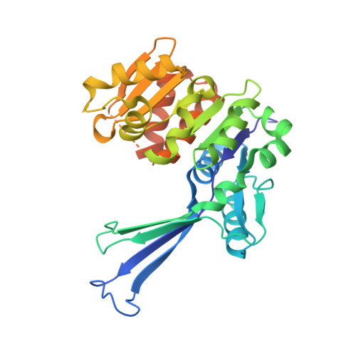
7VSK

Crystal structure of E.coli pseudouridine kinase PsuK complexed with pseudouridine.


Experimental Data Snapshot

  • Method: X-RAY DIFFRACTION
  • Resolution: 2.31 Å
  • R-Value Free: 0.276 
  • R-Value Work: 0.228 
  • R-Value Observed: 0.230 

Starting Model: experimental
View more details

wwPDB Validation   3D Report Full Report


Ligand Structure Quality Assessment 


This is version 1.2 of the entry. See complete history


Literature

Structure Characterization of Escherichia coli Pseudouridine Kinase PsuK.

Li, X.Li, K.Guo, W.Wen, Y.Meng, C.Wu, B.

(2022) Front Microbiol 13: 926099-926099

  • DOI: https://doi.org/10.3389/fmicb.2022.926099
  • Primary Citation of Related Structures:  
    7VKP, 7VSK, 7W93

  • PubMed Abstract: 

    Pseudouridine (Ψ) is one of the most abundant RNA modifications in cellular RNAs that post-transcriptionally impact many aspects of RNA. However, the metabolic fate of modified RNA nucleotides has long been a question. A pseudouridine kinase (PsuK) and a pseudouridine monophosphate glycosylase (PsuG) in Escherichia coli were first characterized as involved in pseudouridine degradation by catalyzing the phosphorylation of pseudouridine to pseudouridine 5'-phosphate (ΨMP) and further hydrolyzing 5'-ΨMP to produce uracil and ribose 5'-phosphate. Recently, their homolog proteins in eukaryotes were also identified, which were named PUKI and PUMY in Arabidopsis . Here, we solved the crystal structures of apo- Ec PsuK and its binary complex with Ψ or N 1 -methyl-pseudouridine (m1Ψ). The structure of Ec PsuK showed a homodimer conformation assembled by its β-thumb region. Ec PsuK has an appropriate binding site with a series of hydrophilic and hydrophobic interactions for Ψ. Moreover, our complex structure of Ec PsuK-m1Ψ suggested the binding pocket has an appropriate capacity for m1Ψ. We also identified the monovalent ion-binding site and potential ATP-binding site. Our studies improved the understanding of the mechanism of Ψ turnover.


  • Organizational Affiliation

    Guangdong Provincial Key Laboratory of Malignant Tumor Epigenetics and Gene Regulation, Guangdong-Hong Kong Joint Laboratory for RNA Medicine, RNA Biomedical Institute, Medical Research Center, Sun Yat-sen Memorial Hospital, Sun Yat-sen University, Guangzhou, China.


Macromolecules
Find similar proteins by:  (by identity cutoff)  |  3D Structure
Entity ID: 1
MoleculeChains Sequence LengthOrganismDetailsImage
PfkB domain protein313Escherichia coli BL21(DE3)Mutation(s): 0 
Gene Names: ECBD_1492
UniProt
Find proteins for A0A140N873 (Escherichia coli (strain B / BL21-DE3))
Explore A0A140N873 
Go to UniProtKB:  A0A140N873
Entity Groups  
Sequence Clusters30% Identity50% Identity70% Identity90% Identity95% Identity100% Identity
UniProt GroupA0A140N873
Sequence Annotations
Expand
  • Reference Sequence
Small Molecules
Ligands 1 Unique
IDChains Name / Formula / InChI Key2D Diagram3D Interactions
FJF (Subject of Investigation/LOI)
Query on FJF

Download Ideal Coordinates CCD File 
B [auth A]5-[(2~{S},3~{R},4~{S},5~{R})-5-(hydroxymethyl)-3,4-bis(oxidanyl)oxolan-2-yl]-1~{H}-pyrimidine-2,4-dione
C9 H12 N2 O6
PTJWIQPHWPFNBW-GBNDHIKLSA-N
Experimental Data & Validation

Experimental Data

  • Method: X-RAY DIFFRACTION
  • Resolution: 2.31 Å
  • R-Value Free: 0.276 
  • R-Value Work: 0.228 
  • R-Value Observed: 0.230 
  • Space Group: P 63 2 2
Unit Cell:
Length ( Å )Angle ( ˚ )
a = 182.634α = 90
b = 182.634β = 90
c = 51.228γ = 120
Software Package:
Software NamePurpose
PHENIXrefinement
HKL-3000data reduction
HKL-3000data scaling
PHASERphasing

Structure Validation

View Full Validation Report


Ligand Structure Quality Assessment 


Entry History & Funding Information

Deposition Data


Funding OrganizationLocationGrant Number
Not funded--

Revision History  (Full details and data files)

  • Version 1.0: 2022-06-22
    Type: Initial release
  • Version 1.1: 2022-07-20
    Changes: Database references
  • Version 1.2: 2023-11-29
    Changes: Data collection, Refinement description