7VIO | pdb_00007vio

Crystal structure of recombinant horse spleen apo-R168H/L169C-Fr


Experimental Data Snapshot

  • Method: X-RAY DIFFRACTION
  • Resolution: 1.50 Å
  • R-Value Free: 
    0.193 (Depositor), 0.227 (DCC) 
  • R-Value Work: 
    0.175 (Depositor) 

Starting Model: experimental
View more details

wwPDB Validation 3D Report Full Report

Validation slider image for 7VIO

This is version 1.2 of the entry. See complete history

Literature

Design of a gold clustering site in an engineered apo-ferritin cage.

Lu, C.Maity, B.Peng, X.Ito, N.Abe, S.Sheng, X.Ueno, T.Lu, D.

(2022) Commun Chem 5: 39-39

  • DOI: https://doi.org/10.1038/s42004-022-00651-1
  • Primary Citation Related Structures: 
    7VIO, 7VIP, 7VIQ, 7VIR, 7VIS, 7VIT, 7VIU

  • PubMed Abstract: 

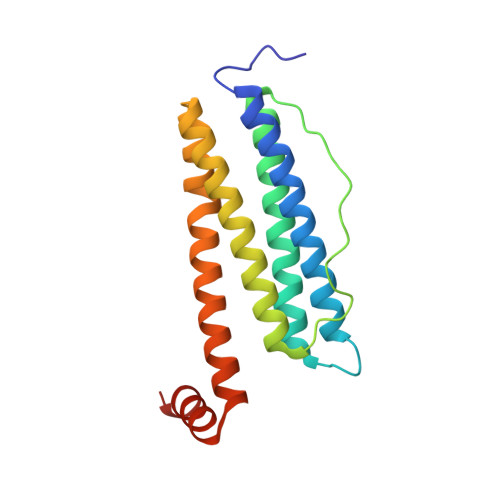
    Water-soluble and biocompatible protein-protected gold nanoclusters (Au NCs) hold great promise for numerous applications. However, design and precise regulation of their structure at an atomic level remain challenging. Herein, we have engineered and constructed a gold clustering site at the 4-fold symmetric axis channel of the apo-ferritin cage. Using a series of X-ray crystal structures, we evaluated the stepwise accumulation process of Au ions into the cage and the formation of a multinuclear Au cluster in our designed cavity. We also disclosed the role of key residues in the metal accumulation process. X-ray crystal structures in combination with quantum chemical (QC) calculation revealed a unique Au clustering site with up to 12 Au atoms positions in the cavity. Moreover, the structure of the gold nanocluster was precisely tuned by the dosage of the Au precursor. As the gold concentration increases, the number of Au atoms position at the clustering site increases from 8 to 12, and a structural rearrangement was observed at a higher Au concentration. Furthermore, the binding affinity order of the four Au binding sites on apo-ferritin was unveiled with a stepwise increase of Au precursor concentration.


  • Organizational Affiliation
    • Department of Chemical Engineering, Tsinghua University, Beijing, 100-084, China.

Macromolecule Content 

  • Total Structure Weight: 20.46 kDa 
  • Atom Count: 1,614 
  • Modeled Residue Count: 171 
  • Deposited Residue Count: 174 
  • Unique protein chains: 1

Macromolecules

Find similar proteins by:|  3D Structure
Entity ID: 1
MoleculeChains  Sequence LengthOrganismDetailsImage
Ferritin light chain174Equus caballusMutation(s): 2 
Gene Names: FTL
UniProt
Find proteins for P02791 (Equus caballus)
Explore P02791 
Go to UniProtKB:  P02791
Entity Groups
Sequence Clusters30% Identity50% Identity70% Identity90% Identity95% Identity100% Identity
UniProt GroupP02791
Sequence Annotations
Expand
Reference Sequence

Experimental Data & Validation

Experimental Data

  • Method: X-RAY DIFFRACTION
  • Resolution: 1.50 Å
  • R-Value Free:  0.193 (Depositor), 0.227 (DCC) 
  • R-Value Work:  0.175 (Depositor) 
Space Group: F 4 3 2
Unit Cell:
Length ( Å )Angle ( ˚ )
a = 182.644α = 90
b = 182.644β = 90
c = 182.644γ = 90
Software Package:
Software NamePurpose
REFMACrefinement
HKL-2000data reduction
Cootmodel building
Aimlessdata scaling
MOLREPphasing

Structure Validation

View Full Validation Report


Entry History 

& Funding Information

Deposition Data


Funding OrganizationLocationGrant Number
National Natural Science Foundation of China (NSFC)China21878175

Revision History  (Full details and data files)

  • Version 1.0: 2022-06-08
    Type: Initial release
  • Version 1.1: 2023-02-08
    Changes: Database references, Derived calculations
  • Version 1.2: 2023-11-29
    Changes: Data collection, Refinement description