7VGR

SARS-CoV-2 M protein dimer (long form) in complex with YN7756_1 Fab


Experimental Data Snapshot

  • Method: ELECTRON MICROSCOPY
  • Resolution: 2.70 Å
  • Aggregation State: PARTICLE 
  • Reconstruction Method: SINGLE PARTICLE 

wwPDB Validation   3D Report Full Report


This is version 1.1 of the entry. See complete history


Literature

Structure of SARS-CoV-2 membrane protein essential for virus assembly.

Zhang, Z.Nomura, N.Muramoto, Y.Ekimoto, T.Uemura, T.Liu, K.Yui, M.Kono, N.Aoki, J.Ikeguchi, M.Noda, T.Iwata, S.Ohto, U.Shimizu, T.

(2022) Nat Commun 13: 4399-4399

  • DOI: https://doi.org/10.1038/s41467-022-32019-3
  • Primary Citation of Related Structures:  
    7VGR, 7VGS

  • PubMed Abstract: 

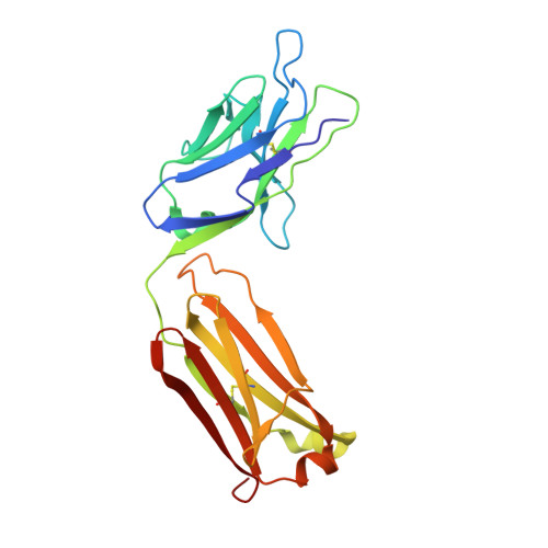
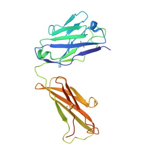
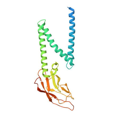
    The coronavirus membrane protein (M) is the most abundant viral structural protein and plays a central role in virus assembly and morphogenesis. However, the process of M protein-driven virus assembly are largely unknown. Here, we report the cryo-electron microscopy structure of the SARS-CoV-2 M protein in two different conformations. M protein forms a mushroom-shaped dimer, composed of two transmembrane domain-swapped three-helix bundles and two intravirion domains. M protein further assembles into higher-order oligomers. A highly conserved hinge region is key for conformational changes. The M protein dimer is unexpectedly similar to SARS-CoV-2 ORF3a, a viral ion channel. Moreover, the interaction analyses of M protein with nucleocapsid protein (N) and RNA suggest that the M protein mediates the concerted recruitment of these components through the positively charged intravirion domain. Our data shed light on the M protein-driven virus assembly mechanism and provide a structural basis for therapeutic intervention targeting M protein.


  • Organizational Affiliation

    Graduate School of Pharmaceutical Sciences, The University of Tokyo, 7-3-1 Hongo, Bunkyo-ku, Tokyo, Japan.


Macromolecules
Find similar proteins by:  (by identity cutoff)  |  3D Structure
Entity ID: 1
MoleculeChains Sequence LengthOrganismDetailsImage
YN7756_1 Fab light chainA [auth C],
C [auth E]
218Mus musculusMutation(s): 0 
Entity Groups  
Sequence Clusters30% Identity50% Identity70% Identity90% Identity95% Identity100% Identity
Sequence Annotations
Expand
  • Reference Sequence
Find similar proteins by:  (by identity cutoff)  |  3D Structure
Entity ID: 2
MoleculeChains Sequence LengthOrganismDetailsImage
YN7756_1 Fab heavy chainB [auth D],
D [auth F]
236Mus musculusMutation(s): 0 
Entity Groups  
Sequence Clusters30% Identity50% Identity70% Identity90% Identity95% Identity100% Identity
Sequence Annotations
Expand
  • Reference Sequence
Find similar proteins by:  (by identity cutoff)  |  3D Structure
Entity ID: 3
MoleculeChains Sequence LengthOrganismDetailsImage
Membrane proteinE [auth A],
F [auth B]
246Severe acute respiratory syndrome coronavirus 2Mutation(s): 0 
Membrane Entity: Yes 
UniProt
Find proteins for P0DTC5 (Severe acute respiratory syndrome coronavirus 2)
Explore P0DTC5 
Go to UniProtKB:  P0DTC5
Entity Groups  
Sequence Clusters30% Identity50% Identity70% Identity90% Identity95% Identity100% Identity
UniProt GroupP0DTC5
Sequence Annotations
Expand
  • Reference Sequence
Experimental Data & Validation

Experimental Data

  • Method: ELECTRON MICROSCOPY
  • Resolution: 2.70 Å
  • Aggregation State: PARTICLE 
  • Reconstruction Method: SINGLE PARTICLE 

Structure Validation

View Full Validation Report


Entry History & Funding Information

Deposition Data


Funding OrganizationLocationGrant Number
Not funded--

Revision History  (Full details and data files)

  • Version 1.0: 2022-08-03
    Type: Initial release
  • Version 1.1: 2023-08-16
    Changes: Data collection, Database references