7VF3

Plexin B1 extracellular fragment in complex with lasso-grafted PB1m7 peptide


Experimental Data Snapshot

  • Method: X-RAY DIFFRACTION
  • Resolution: 2.29 Å
  • R-Value Free: 0.226 
  • R-Value Work: 0.196 
  • R-Value Observed: 0.198 

wwPDB Validation   3D Report Full Report


Ligand Structure Quality Assessment 


This is version 1.3 of the entry. See complete history


Literature

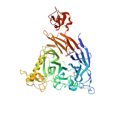
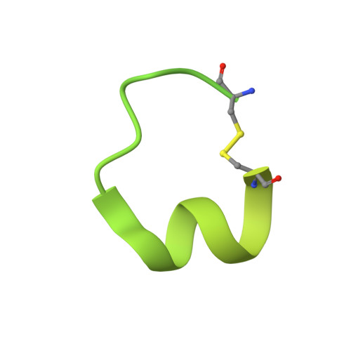
De novo Fc-based receptor dimerizers differentially modulate PlexinB1 function.

Sugano-Nakamura, N.Matoba, K.Hirose, M.Bashiruddin, N.K.Matsunaga, Y.Yamashita, K.Hirata, K.Yamamoto, M.Arimori, T.Suga, H.Takagi, J.

(2022) Structure 30: 1411-1423.e4

  • DOI: https://doi.org/10.1016/j.str.2022.07.008
  • Primary Citation of Related Structures:  
    7VF3, 7VG7

  • PubMed Abstract: 

    Signaling by single-pass transmembrane receptors often involves a formation of ligand-induced receptor dimers with particular conformation, and bivalent receptor binders can modulate receptor functions by inducing different receptor dimer conformations, although such agents are difficult to design. Here, we describe the generation of both antagonistic and agonistic receptor dimerizers toward PlexinB1 (PlxnB1), a receptor for semaphorin 4D (Sema4D), by grafting two different PlxnB1-binding peptides onto the human immunoglobulin G1 (IgG1) Fc protein. The function-modulating activity of a peptide Fc was strongly dependent on the type of the peptide as well as the grafting site, with the best variants showing activity at an nM concentration range. Structural analysis of each peptide-PlxnB1 complex revealed that the agonistic Fc dimerizes PlxnB1 in a face-to-face fashion similar to that induced by Sema4D, whereas antagonistic Fc would induce signaling-incompetent PlxnB1 dimer conformation, enforcing the idea that plexin activation is primarily controlled by the receptor orientation within the dimer.


  • Organizational Affiliation

    Laboratory for Protein Synthesis and Expression, Institute for Protein Research, Osaka University, Osaka, Japan.


Macromolecules
Find similar proteins by:  (by identity cutoff)  |  3D Structure
Entity ID: 1
MoleculeChains Sequence LengthOrganismDetailsImage
Plexin-B1
A, C
525Homo sapiensMutation(s): 1 
Gene Names: PLXNB1KIAA0407PLXN5SEP
UniProt & NIH Common Fund Data Resources
Find proteins for O43157 (Homo sapiens)
Explore O43157 
Go to UniProtKB:  O43157
PHAROS:  O43157
GTEx:  ENSG00000164050 
Entity Groups  
Sequence Clusters30% Identity50% Identity70% Identity90% Identity95% Identity100% Identity
UniProt GroupO43157
Sequence Annotations
Expand
  • Reference Sequence
Find similar proteins by:  (by identity cutoff)  |  3D Structure
Entity ID: 2
MoleculeChains Sequence LengthOrganismDetailsImage
Uteroglobin,PB1m7 peptide,Uteroglobin
B, D
155Homo sapiensMutation(s): 1 
Gene Names: SCGB1A1CC10CCSPUGB
UniProt & NIH Common Fund Data Resources
Find proteins for P11684 (Homo sapiens)
Explore P11684 
Go to UniProtKB:  P11684
PHAROS:  P11684
GTEx:  ENSG00000149021 
Entity Groups  
Sequence Clusters30% Identity50% Identity70% Identity90% Identity95% Identity100% Identity
UniProt GroupP11684
Sequence Annotations
Expand
  • Reference Sequence
Small Molecules
Ligands 4 Unique
IDChains Name / Formula / InChI Key2D Diagram3D Interactions
NAG
Query on NAG

Download Ideal Coordinates CCD File 
E [auth A],
G [auth C]
2-acetamido-2-deoxy-beta-D-glucopyranose
C8 H15 N O6
OVRNDRQMDRJTHS-FMDGEEDCSA-N
PG4
Query on PG4

Download Ideal Coordinates CCD File 
I [auth C],
J [auth C],
K [auth C],
L [auth C]
TETRAETHYLENE GLYCOL
C8 H18 O5
UWHCKJMYHZGTIT-UHFFFAOYSA-N
PGE
Query on PGE

Download Ideal Coordinates CCD File 
H [auth C]TRIETHYLENE GLYCOL
C6 H14 O4
ZIBGPFATKBEMQZ-UHFFFAOYSA-N
PEG
Query on PEG

Download Ideal Coordinates CCD File 
F [auth A]DI(HYDROXYETHYL)ETHER
C4 H10 O3
MTHSVFCYNBDYFN-UHFFFAOYSA-N
Experimental Data & Validation

Experimental Data

  • Method: X-RAY DIFFRACTION
  • Resolution: 2.29 Å
  • R-Value Free: 0.226 
  • R-Value Work: 0.196 
  • R-Value Observed: 0.198 
  • Space Group: I 41 2 2
Unit Cell:
Length ( Å )Angle ( ˚ )
a = 263.591α = 90
b = 263.591β = 90
c = 108.379γ = 90
Software Package:
Software NamePurpose
PHENIXrefinement
XDSdata reduction
XDSdata scaling
PHASERphasing

Structure Validation

View Full Validation Report


Ligand Structure Quality Assessment 


Entry History & Funding Information

Deposition Data


Funding OrganizationLocationGrant Number
Japan Agency for Medical Research and Development (AMED)JapanJP20am0101075

Revision History  (Full details and data files)

  • Version 1.0: 2022-08-17
    Type: Initial release
  • Version 1.1: 2022-08-31
    Changes: Database references
  • Version 1.2: 2022-10-19
    Changes: Database references
  • Version 1.3: 2023-11-29
    Changes: Data collection, Refinement description