Download Mendeley
Crystal structures of the N-terminal and C-terminal domains of the SARS-CoV-2 nucleocapsid protein.
Song, X., Chalhoub, G., McCormick, P.J., Hu, T., Li, J.(2026) Acta Crystallogr F Struct Biol Commun 82: 402-409
- PubMed: 42847964 Search on PubMedSearch on PubMed Central
- DOI: https://doi.org/10.1107/S2053230X26010058
- Primary Citation Related Structures:
7VBD, 7VBE, 7VBF - PubMed Abstract:
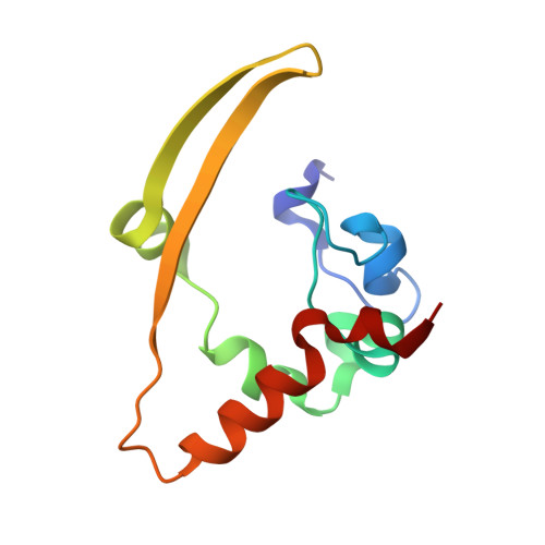
The nucleocapsid (N) protein of Severe acute respiratory syndrome coronavirus 2 (SARS-CoV-2) functions in viral RNA binding, genome packaging and ribonucleoprotein (RNP) assembly. Crystal structures were determined of the N-terminal domain (NTD) from crystals grown at pH 8.0 to a resolution of 1.94 Å and of the C-terminal domain (CTD) from crystals grown at pH 5.0 and 8.5 to resolutions of 1.59 and 1.30 Å, respectively. The NTD adopts the canonical coronavirus NTD fold and contains a positively charged cleft corresponding to a previously proposed RNA-binding region. Comparison of the apo NTD structure determined in this study with a previously reported RNA-bound NTD structure revealed local differences in the side-chain conformations of RNA-contacting residues, including Thr49, Arg88, Arg92, Arg107, Tyr109 and Tyr111, whereas the overall domain fold remained essentially unchanged. These observations suggest that RNA binding may be accommodated by local side-chain rearrangements within a pre-existing positively charged cleft rather than by a large-scale conformational change. The CTD forms a domain-swapped dimer mediated by β-hairpins, with a positively charged groove extending across the dimer interface. Superposition of the two CTD structures yielded a root-mean-square deviation (r.m.s.d.) of 0.129 Å, and 11 intersubunit hydrogen bonds were observed at the dimer interface in each structure, indicating that the dimer architecture is conserved under the crystallization conditions examined. Together, these structures provide a framework for further investigation of NTD-RNA recognition, the possible role of the CTD positively charged groove in nucleic acid interactions and N-protein assembly.
- Key Laboratory of Infection and Immunity, Health Commission of Jiangxi Province and School of Basic Medicine, Nanchang Medical College, Nanchang 330052, People's Republic of China.
Organizational Affiliation: