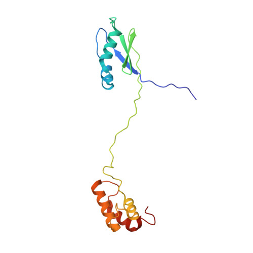
The flexible N-terminal motif of uL11 unique to eukaryotic ribosomes interacts with P-complex and facilitates protein translation.
Yang, L., Lee, K.M., Yu, C.W., Imai, H., Choi, A.K., Banfield, D.K., Ito, K., Uchiumi, T., Wong, K.B.(2022) Nucleic Acids Res 50: 5335-5348
- PubMed: 35544198 Search on PubMedSearch on PubMed Central
- DOI: https://doi.org/10.1093/nar/gkac292
- Primary Citation Related Structures:
7VB2 - PubMed Abstract:
Eukaryotic uL11 contains a conserved MPPKFDP motif at the N-terminus that is not found in archaeal and bacterial homologs. Here, we determined the solution structure of human uL11 by NMR spectroscopy and characterized its backbone dynamics by 15N-1H relaxation experiments. We showed that these N-terminal residues are unstructured and flexible. Structural comparison with ribosome-bound uL11 suggests that the linker region between the N-terminal domain and C-terminal domain of human uL11 is intrinsically disordered and only becomes structured when bound to the ribosomes. Mutagenesis studies show that the N-terminal conserved MPPKFDP motif is involved in interacting with the P-complex and its extended protuberant domain of uL10 in vitro. Truncation of the MPPKFDP motif also reduced the poly-phenylalanine synthesis in both hybrid ribosome and yeast mutagenesis studies. In addition, G→A/P substitutions to the conserved GPLG motif of helix-1 reduced poly-phenylalanine synthesis to 9-32% in yeast ribosomes. We propose that the flexible N-terminal residues of uL11, which could extend up to ∼25 Å from the N-terminal domain of uL11, can form transient interactions with the uL10 that help to fetch and fix it into a position ready for recruiting the incoming translation factors and facilitate protein synthesis.
- School of Life Sciences, Centre for Protein Science and Crystallography, State Key Laboratory of Agrobiotechnology, The Chinese University of Hong Kong, Shatin, Hong Kong, China.
Organizational Affiliation: