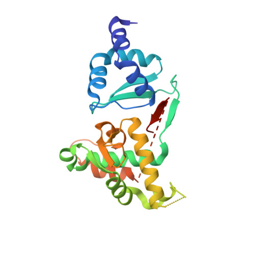
Crystal structure of the chromosome partition protein MukE homodimer.
Qian, J.W., Wang, X.Y., Deng, K., Li, D.F., Guo, L.(2021) Biochem Biophys Res Commun 589: 229-233
- PubMed: 34929446 Search on PubMed
- DOI: https://doi.org/10.1016/j.bbrc.2021.12.032
- Primary Citation Related Structures:
7V8P - PubMed Abstract:
The SMC (structural maintenance of chromosomes) proteins are known to be involved in chromosome pairing or aggregation and play an important role in cell cycle and division. Different from SMC-ScpAB complex maintaining chromosome structure in most bacteria, the MukB-MukE-MukF complex is responsible for chromosome condensation in E. coli and some γ-proteobacter. Though different models were proposed to illustrate the mechanism of how the MukBEF complex worked, the assembly of the MukBEF complex is a key. The MukE dimer interacted with the middle region of one MukF molecule, and was clamped by the N- and C-terminal domain of the latter, and then was involved in the interaction with the head domain of MukB. To reveal the structural basis of MukE involved in the dynamic equilibrium of potential different MukBEF assemblies, we determined the MukE structure at 2.44 Å resolution. We found that the binding cavity for the α10, β4 and β5 of MukF (residues 296-327) in the MukE dimer has been occupied by the α9 and β7 strand of MukE. We proposed that the highly dynamic C-terminal region (173-225) was important for the MukE-F assembly and then involved in the MukBEF complex formation.
- State Key Laboratory of Microbial Resources, Institute of Microbiology, Chinese Academy of Sciences, Beijing, 100101, China; College of Life and Health Sciences, Northeastern University, Shenyang, 110169, China.
Organizational Affiliation: