7V6M | pdb_00007v6m

Crystal structure of lacto-N-biosidase TnX from Tynzenella nexilis, lacto-N-biose complex


Experimental Data Snapshot

  • Method: X-RAY DIFFRACTION
  • Resolution: 1.92 Å
  • R-Value Free: 
    0.209 (Depositor), 0.219 (DCC) 
  • R-Value Work: 
    0.170 (Depositor), 0.181 (DCC) 
  • R-Value Observed: 
    0.172 (Depositor) 

Starting Model: experimental
View more details

wwPDB Validation 3D Report Full Report

Validation slider image for 7V6M

This is version 1.2 of the entry. See complete history

Literature

Crystal structures of glycoside hydrolase family 136 lacto-N-biosidases from monkey gut- and human adult gut bacteria.

Yamada, C.Katayama, T.Fushinobu, S.

(2022) Biosci Biotechnol Biochem 86: 464-475

  • DOI: https://doi.org/10.1093/bbb/zbac015
  • Primary Citation Related Structures: 
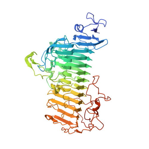
    7V6I, 7V6M

  • PubMed Abstract: 

    Glycoside hydrolase family 136 (GH136) was established after the discovery and structural analysis of lacto-N-biosidase (LNBase) from the infant gut bacterium Bifidobacterium longum subsp. longum JCM1217 (BlLnbX). Homologous genes of BlLnbX are widely distributed in the genomes of human gut bacteria and monkey Bifidobacterium spp., although only 2 crystal structures were reported in the GH136 family. Cell suspensions of Bifidobacterium saguini, Tyzzerella nexilis, and Ruminococcus lactaris exhibited the LNBase activity. Recombinant LNBases of these 3 species were functionally expressed with their specific chaperones in Escherichia coli, and their kinetic parameters against p-nitrophenol substrates were determined. The crystal structures of the LNBases from B. saguini and T. nexilis in complex with lacto-N-biose I were determined at 2.51 and 1.92 Å resolutions, respectively. These structures conserve a β-helix fold characteristic of GH136 and the catalytic residues, but they lack the metal ions that were present in BlLnbX.


  • Organizational Affiliation
    • Department of Biotechnology, Graduate School of Agricultural and Life Sciences, The University of Tokyo, Tokyo, Japan.

Macromolecule Content 

  • Total Structure Weight: 65.25 kDa 
  • Atom Count: 4,856 
  • Modeled Residue Count: 570 
  • Deposited Residue Count: 587 
  • Unique protein chains: 1

Macromolecules

Find similar proteins by:|  3D Structure
Entity ID: 1
MoleculeChains  Sequence LengthOrganismDetailsImage
Fibronectin type III domain-containing protein587Faecalimonas nexilisMutation(s): 0 
Gene Names: DXA47_02055

Oligosaccharides

Help  
Entity ID: 2
MoleculeChains Length2D Diagram GlycosylationD Interactions
beta-D-galactopyranose-(1-3)-2-acetamido-2-deoxy-beta-D-glucopyranose
B
2N/A
Glycosylation Resources
GlyTouCan: G00056MO
GlyCosmos: G00056MO
GlyGen: G00056MO

Experimental Data & Validation

Experimental Data

  • Method: X-RAY DIFFRACTION
  • Resolution: 1.92 Å
  • R-Value Free:  0.209 (Depositor), 0.219 (DCC) 
  • R-Value Work:  0.170 (Depositor), 0.181 (DCC) 
  • R-Value Observed: 0.172 (Depositor) 
Space Group: P 2 21 21
Unit Cell:
Length ( Å )Angle ( ˚ )
a = 55.205α = 90
b = 71.782β = 90
c = 161.488γ = 90
Software Package:
Software NamePurpose
REFMACrefinement
XDSdata reduction
Aimlessdata scaling
MOLREPphasing

Structure Validation

View Full Validation Report


Entry History 

& Funding Information

Deposition Data


Funding OrganizationLocationGrant Number
Japan Society for the Promotion of Science (JSPS)Japan16J09251
Japan Society for the Promotion of Science (JSPS)Japan18K14388

Revision History  (Full details and data files)

  • Version 1.0: 2022-02-09
    Type: Initial release
  • Version 1.1: 2022-04-06
    Changes: Database references
  • Version 1.2: 2023-11-29
    Changes: Data collection, Refinement description