7V4E | pdb_00007v4e

Crystal Structure of VpsR display novel dimeric architecture and c-di-GMP binding: mechanistic implications in oligomerization, ATPase activity and DNA binding.


Experimental Data Snapshot

  • Method: X-RAY DIFFRACTION
  • Resolution: 4.00 Å
  • R-Value Free: 
    0.280 (Depositor), 0.294 (DCC) 
  • R-Value Work: 
    0.241 (Depositor), 0.260 (DCC) 
  • R-Value Observed: 
    0.245 (Depositor) 

Starting Model: experimental
View more details

wwPDB Validation 3D Report Full Report

Validation slider image for 7V4E

Ligand Structure Quality Assessment 


This is version 1.1 of the entry. See complete history

Literature

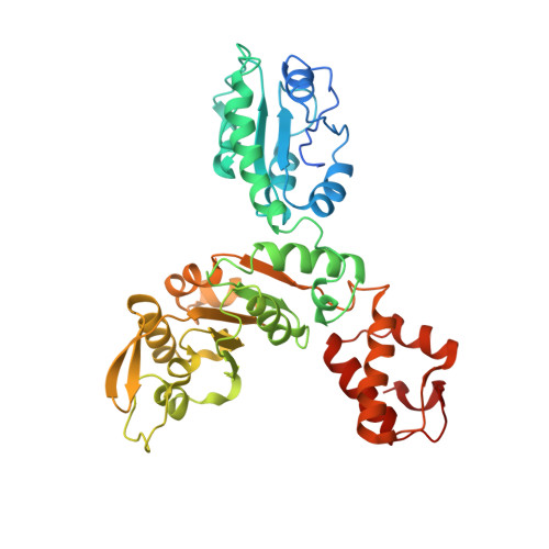
Crystal Structure of VpsR Revealed Novel Dimeric Architecture and c-di-GMP Binding Site: Mechanistic Implications in Oligomerization, ATPase Activity and DNA Binding.

Chakrabortty, T.Roy Chowdhury, S.Ghosh, B.Sen, U.

(2022) J Mol Biology 434: 167354-167354

  • DOI: https://doi.org/10.1016/j.jmb.2021.167354
  • Primary Citation Related Structures: 
    7V2B, 7V2V, 7V3W, 7V4E

  • PubMed Abstract: 

    VpsR, the master regulator of biofilm formation in Vibrio cholerae, is an atypical NtrC1 type bEBP lacking residues essential for σ 54 -RNAP binding and REC domain phosphorylation. Moreover, transcription from P vpsL , a promoter of biofilm biosynthesis, has been documented in presence of σ 70 -RNAP/VpsR/c-di-GMP complex. It was proposed that c-di-GMP and VpsR together form an active transcription complex with σ 70 -RNAP. However, the impact of c-di-GMP imparted on VpsR that leads to transcription activation with σ 70 -RNAP remained elusive, largely due to the lack of the structure of VpsR and knowledge about c-di-GMP:VpsR interactions. In this direction we have solved the crystal structure of VpsR RA , containing REC and AAA + domains, in apo, AMPPNP/GMPPNP and c-di-GMP bound states. Structures of VpsR RA unveiled distinctive REC domain orientation that leads to a novel dimeric association and noncanonical ATP/GTP binding. Moreover, we have demonstrated that at physiological pH VpsR remains as monomer having no ATPase activity but c-di-GMP imparted cooperativity to convert it to dimer with potent activity. Crystal structure of c-di-GMP:VpsR RA complex reveals that c-di-GMP binds near the C-terminal end of AAA + domain. Trp quenching studies on VpsR R , VpsR A , VpsR RA , VpsR AD with c-di-GMP additionally demonstrated that c-di-GMP could potentially bind VpsR D . We propose that c-di-GMP mediated tethering of VpsR D with VpsR A could likely favor generating the specific protein-DNA architecture for transcription activation.


  • Organizational Affiliation
    • Crystallography and Molecular Biology Division, Saha Institute of Nuclear Physics, HBNI, 1/AF Bidhan Nagar, Kolkata 700064, India. Electronic address: https://twitter.com/@TulikaC02382598.

Macromolecule Content 

  • Total Structure Weight: 91.02 kDa 
  • Atom Count: 5,974 
  • Modeled Residue Count: 748 
  • Deposited Residue Count: 798 
  • Unique protein chains: 1

Macromolecules

Find similar proteins by:|  3D Structure
Entity ID: 1
MoleculeChains  Sequence LengthOrganismDetailsImage
VpsRA,
B [auth D]
399Vibrio choleraeMutation(s): 0 
Gene Names: vpsR
UniProt
Find proteins for Q9KU59 (Vibrio cholerae serotype O1 (strain ATCC 39315 / El Tor Inaba N16961))
Explore Q9KU59 
Go to UniProtKB:  Q9KU59
Entity Groups
Sequence Clusters30% Identity50% Identity70% Identity90% Identity95% Identity100% Identity
UniProt GroupQ9KU59
Sequence Annotations
Expand
Reference Sequence

Small Molecules

Ligands 2 Unique
IDChains Name / Formula / InChI Key2D Diagram3D Interactions
C2E
(Subject of Investigation/LOI)

Query on C2E


Download:Ideal Coordinates CCD File
E [auth D],
F [auth D]
9,9'-[(2R,3R,3aS,5S,7aR,9R,10R,10aS,12S,14aR)-3,5,10,12-tetrahydroxy-5,12-dioxidooctahydro-2H,7H-difuro[3,2-d:3',2'-j][1,3,7,9,2,8]tetraoxadiphosphacyclododecine-2,9-diyl]bis(2-amino-1,9-dihydro-6H-purin-6-one)
C20 H24 N10 O14 P2
PKFDLKSEZWEFGL-MHARETSRSA-N
SO4
(Subject of Investigation/LOI)

Query on SO4


Download:Ideal Coordinates CCD File
C [auth A],
D
SULFATE ION
O4 S
QAOWNCQODCNURD-UHFFFAOYSA-L

Experimental Data & Validation

Experimental Data

  • Method: X-RAY DIFFRACTION
  • Resolution: 4.00 Å
  • R-Value Free:  0.280 (Depositor), 0.294 (DCC) 
  • R-Value Work:  0.241 (Depositor), 0.260 (DCC) 
  • R-Value Observed: 0.245 (Depositor) 
Space Group: P 31
Unit Cell:
Length ( Å )Angle ( ˚ )
a = 119.687α = 90
b = 119.687β = 90
c = 81.141γ = 120
Software Package:
Software NamePurpose
PHENIXrefinement
XDSdata reduction
Aimlessdata scaling
PHASERphasing

Structure Validation

View Full Validation Report


Ligand Structure Quality Assessment 


Entry History 

& Funding Information

Deposition Data


Funding OrganizationLocationGrant Number
Department of Science & Technology (DST, India)IndiaEMR/2015/000780 Dated 18-07-2016

Revision History  (Full details and data files)

  • Version 1.0: 2022-04-06
    Type: Initial release
  • Version 1.1: 2023-11-29
    Changes: Data collection, Refinement description