


Crystal structure of SULT3A1, mouse amine N-sulfotransferase
Teramoto, T., Inada, K., Kurogi, K., Sakakibara, Y., Kakuta, Y.To be published.
Experimental Data Snapshot
Entity ID: 1 | |||||
|---|---|---|---|---|---|
| Molecule | Chains | Sequence Length | Organism | Details | Image |
| Amine sulfotransferase | 293 | Mus musculus | Mutation(s): 2 Gene Names: Sult3a1, St3a1 EC: 2.8.2.3 | 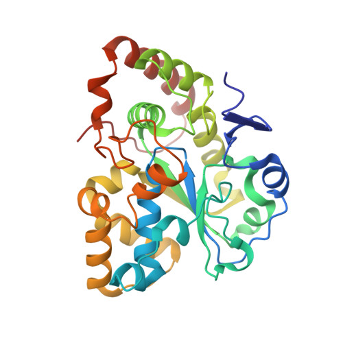 | |
UniProt | |||||
Entity Groups | |||||
| Sequence Clusters | 30% Identity50% Identity70% Identity90% Identity95% Identity100% Identity | ||||
| UniProt Group | O35403 | ||||
Sequence AnnotationsExpand | |||||
Reference Sequence | |||||
| Ligands 2 Unique | |||||
|---|---|---|---|---|---|
| ID | Chains | Name / Formula / InChI Key | 2D Diagram | 3D Interactions | |
| A3P (Subject of Investigation/LOI) Download:Ideal Coordinates CCD File | D [auth A], F [auth B] | ADENOSINE-3'-5'-DIPHOSPHATE C10 H15 N5 O10 P2 WHTCPDAXWFLDIH-KQYNXXCUSA-N |  | ||
| 5I2 (Subject of Investigation/LOI) Download:Ideal Coordinates CCD File | C [auth A], E [auth B] | naphthalen-1-amine C10 H9 N RUFPHBVGCFYCNW-UHFFFAOYSA-N |  | ||
| Length ( Å ) | Angle ( ˚ ) |
|---|---|
| a = 116.44 | α = 90 |
| b = 116.44 | β = 90 |
| c = 203.31 | γ = 120 |
| Software Name | Purpose |
|---|---|
| PHENIX | refinement |
| HKL-2000 | data reduction |
| HKL-2000 | data scaling |
| SOLVE | phasing |
| RESOLVE | phasing |
| Funding Organization | Location | Grant Number |
|---|---|---|
| Japan Society for the Promotion of Science (JSPS) | Japan | 21K05384 |


