7UTJ

Cryogenic electron microscopy 3D map of F-actin bound by human dimeric alpha-catenin


Experimental Data Snapshot

  • Method: ELECTRON MICROSCOPY
  • Resolution: 2.77 Å
  • Aggregation State: FILAMENT 
  • Reconstruction Method: HELICAL 

wwPDB Validation   3D Report Full Report


This is version 1.1 of the entry. See complete history


Literature

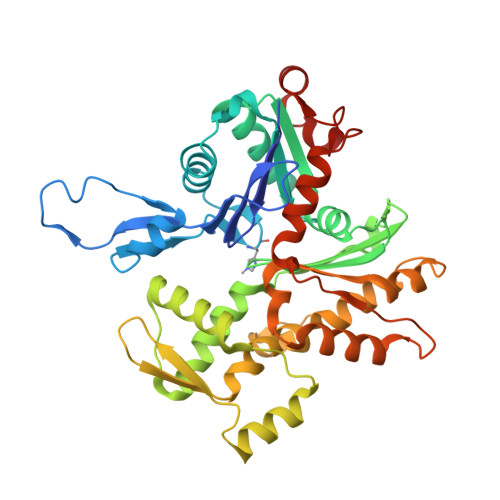
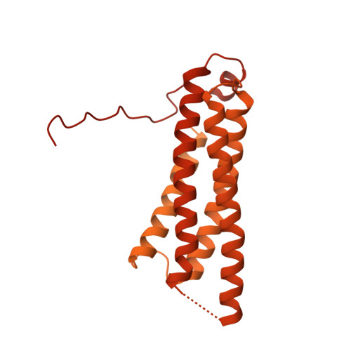
Distinct inter-domain interactions of dimeric versus monomeric alpha-catenin link cell junctions to filaments.

Rangarajan, E.S.Smith, E.W.Izard, T.

(2023) Commun Biol 6: 276-276

  • DOI: https://doi.org/10.1038/s42003-023-04610-x
  • Primary Citation of Related Structures:  
    7UTJ, 7UXF

  • PubMed Abstract: 

    Attachment between cells is crucial for almost all aspects of the life of cells. These inter-cell adhesions are mediated by the binding of transmembrane cadherin receptors of one cell to cadherins of a neighboring cell. Inside the cell, cadherin binds β-catenin, which interacts with α-catenin. The transitioning of cells between migration and adhesion is modulated by α-catenin, which links cell junctions and the plasma membrane to the actin cytoskeleton. At cell junctions, a single β-catenin/α-catenin heterodimer slips along filamentous actin in the direction of cytoskeletal tension which unfolds clustered heterodimers to form catch bonds with F-actin. Outside cell junctions, α-catenin dimerizes and links the plasma membrane to F-actin. Under cytoskeletal tension, α-catenin unfolds and forms an asymmetric catch bond with F-actin. To understand the mechanism of this important α-catenin function, we determined the 2.7 Å cryogenic electron microscopy (cryoEM) structures of filamentous actin alone and bound to human dimeric α-catenin. Our structures provide mechanistic insights into the role of the α-catenin interdomain interactions in directing α-catenin function and suggest a bivalent mechanism. Further, our cryoEM structure of human monomeric α-catenin provides mechanistic insights into α-catenin autoinhibition. Collectively, our structures capture the initial α-catenin interaction with F-actin before the sensing of force, which is a crucial event in cell adhesion and human disease.


  • Organizational Affiliation

    The Cell Adhesion Laboratory, UF Scripps, Jupiter, FL, 33458, USA.


Macromolecules
Find similar proteins by:  (by identity cutoff)  |  3D Structure
Entity ID: 1
MoleculeChains Sequence LengthOrganismDetailsImage
Actin, alpha skeletal muscle
A, B, C, D, E
A, B, C, D, E, F
377Oryctolagus cuniculusMutation(s): 0 
Gene Names: ACTA1ACTA
UniProt
Find proteins for P68135 (Oryctolagus cuniculus)
Explore P68135 
Go to UniProtKB:  P68135
Entity Groups  
Sequence Clusters30% Identity50% Identity70% Identity90% Identity95% Identity100% Identity
UniProt GroupP68135
Sequence Annotations
Expand
  • Reference Sequence
Find similar proteins by:  (by identity cutoff)  |  3D Structure
Entity ID: 2
MoleculeChains Sequence LengthOrganismDetailsImage
Catenin alpha-1889Homo sapiensMutation(s): 0 
Gene Names: CTNNA1
UniProt & NIH Common Fund Data Resources
Find proteins for P35221 (Homo sapiens)
Explore P35221 
Go to UniProtKB:  P35221
PHAROS:  P35221
GTEx:  ENSG00000044115 
Entity Groups  
Sequence Clusters30% Identity50% Identity70% Identity90% Identity95% Identity100% Identity
UniProt GroupP35221
Sequence Annotations
Expand
  • Reference Sequence
Small Molecules
Ligands 2 Unique
IDChains Name / Formula / InChI Key2D Diagram3D Interactions
ADP
Query on ADP

Download Ideal Coordinates CCD File 
M [auth A]
O [auth B]
Q [auth C]
S [auth D]
U [auth E]
M [auth A],
O [auth B],
Q [auth C],
S [auth D],
U [auth E],
W [auth F]
ADENOSINE-5'-DIPHOSPHATE
C10 H15 N5 O10 P2
XTWYTFMLZFPYCI-KQYNXXCUSA-N
MG
Query on MG

Download Ideal Coordinates CCD File 
N [auth A]
P [auth B]
R [auth C]
T [auth D]
V [auth E]
N [auth A],
P [auth B],
R [auth C],
T [auth D],
V [auth E],
X [auth F]
MAGNESIUM ION
Mg
JLVVSXFLKOJNIY-UHFFFAOYSA-N
Modified Residues  1 Unique
IDChains TypeFormula2D DiagramParent
HIC
Query on HIC
A, B, C, D, E
A, B, C, D, E, F
L-PEPTIDE LINKINGC7 H11 N3 O2HIS
Experimental Data & Validation

Experimental Data

  • Method: ELECTRON MICROSCOPY
  • Resolution: 2.77 Å
  • Aggregation State: FILAMENT 
  • Reconstruction Method: HELICAL 
EM Software:
TaskSoftware PackageVersion
MODEL REFINEMENTCoot
MODEL REFINEMENTPHENIX
RECONSTRUCTIONcryoSPARC

Structure Validation

View Full Validation Report


Entry History & Funding Information

Deposition Data


Funding OrganizationLocationGrant Number
National Science Foundation (NSF, United States)United States--

Revision History  (Full details and data files)

  • Version 1.0: 2023-03-08
    Type: Initial release
  • Version 1.1: 2023-03-29
    Changes: Database references