7UPN

Maedi visna virus Vif in complex with CypA and E3 ubiquitin ligase


Experimental Data Snapshot

  • Method: ELECTRON MICROSCOPY
  • Resolution: 3.50 Å
  • Aggregation State: PARTICLE 
  • Reconstruction Method: SINGLE PARTICLE 

wwPDB Validation   3D Report Full Report


This is version 1.0 of the entry. See complete history


Literature

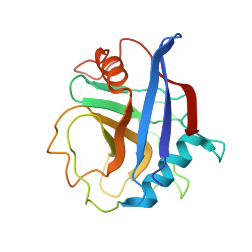
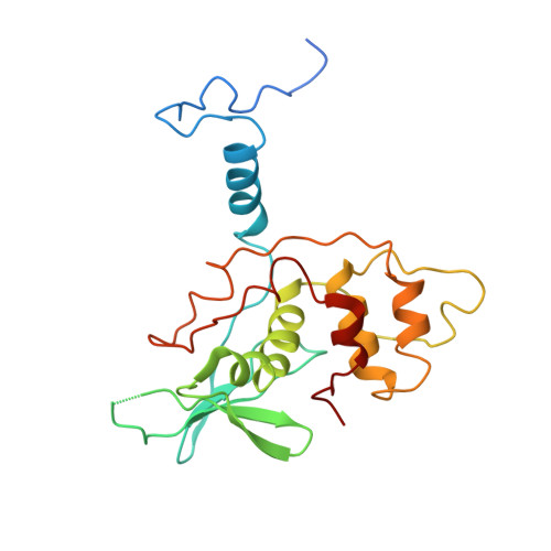
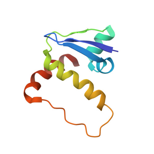
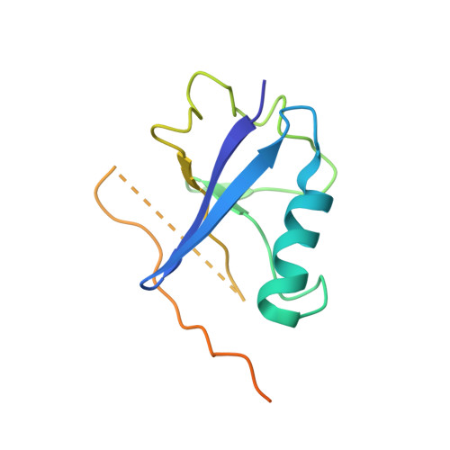
Structural basis for recruitment of host CypA and E3 ubiquitin ligase by maedi-visna virus Vif.

Hu, Y.Gudnadottir, R.B.Knecht, K.M.Arizaga, F.Jonsson, S.R.Xiong, Y.

(2023) Sci Adv 9: eadd3422-eadd3422

  • DOI: https://doi.org/10.1126/sciadv.add3422
  • Primary Citation of Related Structures:  
    7UPN

  • PubMed Abstract: 

    Lentiviral Vif molecules target the host antiviral APOBEC3 proteins for destruction in cellular ubiquitin-proteasome pathways. Different lentiviral Vifs have evolved to use the same canonical E3 ubiquitin ligase complexes, along with distinct noncanonical host cofactors for their activities. Unlike primate lentiviral Vif, which recruits CBFβ as the noncanonical cofactor, nonprimate lentiviral Vif proteins have developed different cofactor recruitment mechanisms. Maedi-visna virus (MVV) sequesters CypA as the noncanonical cofactor for the Vif-mediated ubiquitination of ovine APOBEC3s. Here, we report the cryo-electron microscopy structure of MVV Vif in complex with CypA and E3 ligase components. The structure, along with our biochemical and functional analysis, reveals both conserved and unique structural elements of MVV Vif and its common and distinct interaction modes with various cognate cellular proteins, providing a further understanding of the evolutionary relationship between lentiviral Vifs and the molecular mechanisms by which they capture different host cofactors for immune evasion activities.


  • Organizational Affiliation

    Department of Molecular Biophysics and Biochemistry, Yale University, New Haven, CT, USA.


Macromolecules
Find similar proteins by:  (by identity cutoff)  |  3D Structure
Entity ID: 1
MoleculeChains Sequence LengthOrganismDetailsImage
Peptidyl-prolyl cis-trans isomerase AA,
E [auth B]
165Homo sapiensMutation(s): 0 
Gene Names: PPIACYPA
EC: 5.2.1.8
UniProt & NIH Common Fund Data Resources
Find proteins for P62937 (Homo sapiens)
Explore P62937 
Go to UniProtKB:  P62937
PHAROS:  P62937
GTEx:  ENSG00000196262 
Entity Groups  
Sequence Clusters30% Identity50% Identity70% Identity90% Identity95% Identity100% Identity
UniProt GroupP62937
Sequence Annotations
Expand
  • Reference Sequence
Find similar proteins by:  (by identity cutoff)  |  3D Structure
Entity ID: 2
MoleculeChains Sequence LengthOrganismDetailsImage
Virion infectivity factorB [auth C],
F
230Visna-maedi virusMutation(s): 0 
Gene Names: vif
UniProt
Find proteins for P69717 (Maedi visna virus (strain KV1772))
Explore P69717 
Go to UniProtKB:  P69717
Entity Groups  
Sequence Clusters30% Identity50% Identity70% Identity90% Identity95% Identity100% Identity
UniProt GroupP69717
Sequence Annotations
Expand
  • Reference Sequence
Find similar proteins by:  (by identity cutoff)  |  3D Structure
Entity ID: 3
MoleculeChains Sequence LengthOrganismDetailsImage
Elongin-CC [auth E],
G
96Homo sapiensMutation(s): 0 
Gene Names: ELOCTCEB1
UniProt & NIH Common Fund Data Resources
Find proteins for Q15369 (Homo sapiens)
Explore Q15369 
Go to UniProtKB:  Q15369
PHAROS:  Q15369
GTEx:  ENSG00000154582 
Entity Groups  
Sequence Clusters30% Identity50% Identity70% Identity90% Identity95% Identity100% Identity
UniProt GroupQ15369
Sequence Annotations
Expand
  • Reference Sequence
Find similar proteins by:  (by identity cutoff)  |  3D Structure
Entity ID: 4
MoleculeChains Sequence LengthOrganismDetailsImage
Elongin-BD [auth H],
H [auth I]
118Homo sapiensMutation(s): 0 
Gene Names: ELOBTCEB2
UniProt & NIH Common Fund Data Resources
Find proteins for Q15370 (Homo sapiens)
Explore Q15370 
Go to UniProtKB:  Q15370
PHAROS:  Q15370
GTEx:  ENSG00000103363 
Entity Groups  
Sequence Clusters30% Identity50% Identity70% Identity90% Identity95% Identity100% Identity
UniProt GroupQ15370
Sequence Annotations
Expand
  • Reference Sequence
Experimental Data & Validation

Experimental Data

  • Method: ELECTRON MICROSCOPY
  • Resolution: 3.50 Å
  • Aggregation State: PARTICLE 
  • Reconstruction Method: SINGLE PARTICLE 

Structure Validation

View Full Validation Report


Entry History & Funding Information

Deposition Data


Funding OrganizationLocationGrant Number
National Institutes of Health/National Cancer Institute (NIH/NCI)United States--

Revision History  (Full details and data files)

  • Version 1.0: 2023-01-25
    Type: Initial release