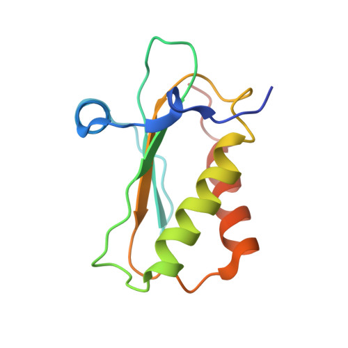
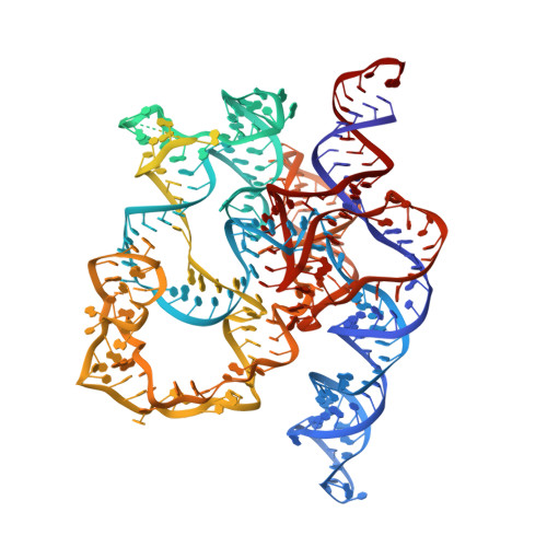
Structural and mechanistic basis for recognition of alternative tRNA precursor substrates by bacterial ribonuclease P.
Zhu, J., Huang, W., Zhao, J., Huynh, L., Taylor, D.J., Harris, M.E.(2022) Nat Commun 13: 5120-5120
- PubMed: 36045135 Search on PubMedSearch on PubMed Central
- DOI: https://doi.org/10.1038/s41467-022-32843-7
- Primary Citation Related Structures:
7UO0, 7UO1, 7UO2, 7UO5 - PubMed Abstract:
Binding of precursor tRNAs (ptRNAs) by bacterial ribonuclease P (RNase P) involves an encounter complex (ES) that isomerizes to a catalytic conformation (ES*). However, the structures of intermediates and the conformational changes that occur during binding are poorly understood. Here, we show that pairing between the 5' leader and 3'RCCA extending the acceptor stem of ptRNA inhibits ES* formation. Cryo-electron microscopy single particle analysis reveals a dynamic enzyme that becomes ordered upon formation of ES* in which extended acceptor stem pairing is unwound. Comparisons of structures with alternative ptRNAs reveals that once unwinding is completed RNase P primarily uses stacking interactions and shape complementarity to accommodate alternative sequences at its cleavage site. Our study reveals active site interactions and conformational changes that drive molecular recognition by RNase P and lays the foundation for understanding how binding interactions are linked to helix unwinding and catalysis.
- Department of Chemistry, University of Florida, Gainesville, FL, USA.
Organizational Affiliation: