7UMS

Structure of the VP5*/VP8* assembly from the human rotavirus strain CDC-9 in complex with antibody 41 - Upright conformation


Experimental Data Snapshot

  • Method: ELECTRON MICROSCOPY
  • Resolution: 3.50 Å
  • Aggregation State: PARTICLE 
  • Reconstruction Method: SINGLE PARTICLE 

wwPDB Validation   3D Report Full Report


This is version 1.3 of the entry. See complete history


Literature

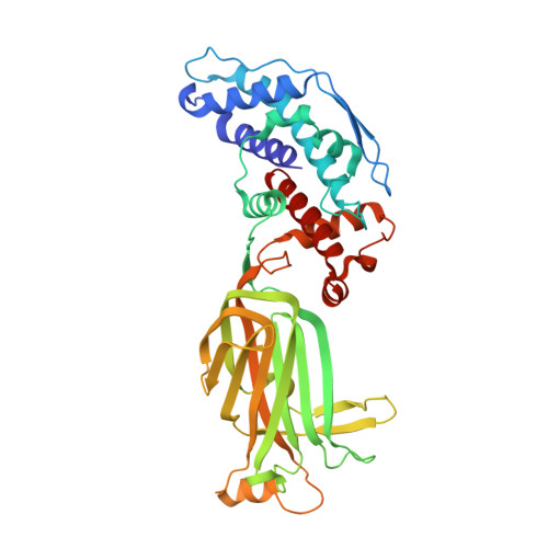
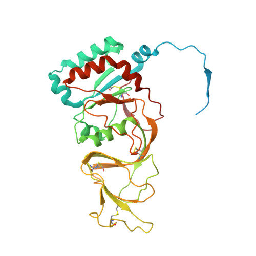
Rotavirus VP4 Epitope of a Broadly Neutralizing Human Antibody Defined by Its Structure Bound with an Attenuated-Strain Virion.

Jenni, S.Li, Z.Wang, Y.Bessey, T.Salgado, E.N.Schmidt, A.G.Greenberg, H.B.Jiang, B.Harrison, S.C.

(2022) J Virol 96: e0062722-e0062722

  • DOI: https://doi.org/10.1128/jvi.00627-22
  • Primary Citation of Related Structures:  
    7UMS, 7UMT

  • PubMed Abstract: 

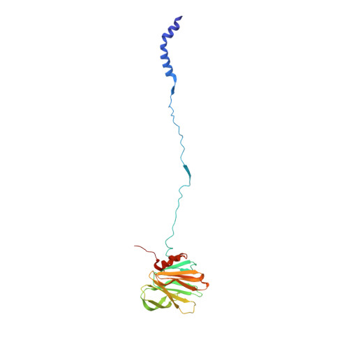
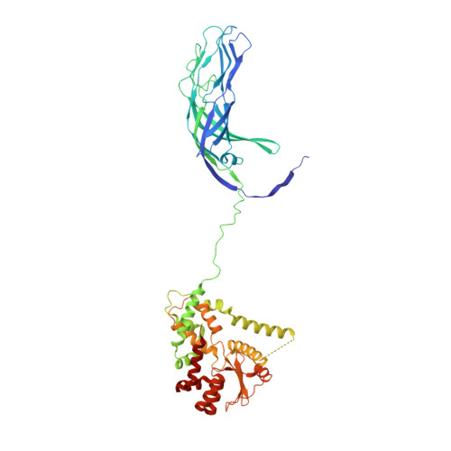
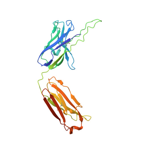
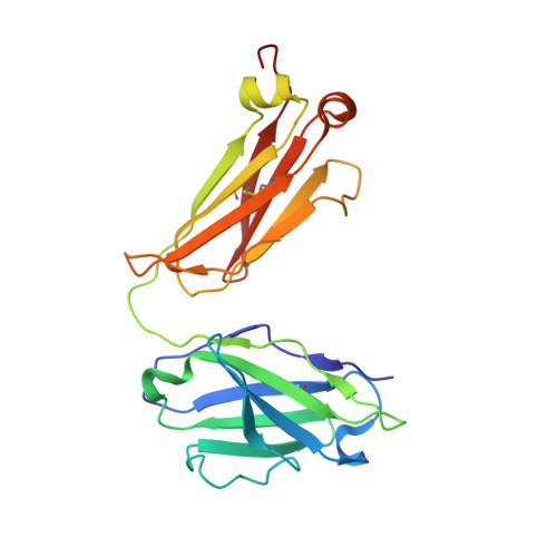
    Rotavirus live-attenuated vaccines, both mono- and pentavalent, generate broadly heterotypic protection. B-cells isolated from adults encode neutralizing antibodies, some with affinity for VP5*, that afford broad protection in mice. We have mapped the epitope of one such antibody by determining the high-resolution cryo-EM structure of its antigen-binding fragment (Fab) bound to the virion of a candidate vaccine strain, CDC-9. The Fab contacts both the distal end of a VP5* β-barrel domain and the two VP8* lectin-like domains at the tip of a projecting spike. Its interactions with VP8* do not impinge on the likely receptor-binding site, suggesting that the mechanism of neutralization is at a step subsequent to initial attachment. We also examined structures of CDC-9 virions from two different stages of serial passaging. Nearly all the VP4 (cleaved to VP8*/VP5*) spikes on particles from the earlier passage (wild-type isolate) had transitioned from the "upright" conformation present on fully infectious virions to the "reversed" conformation that is probably the end state of membrane insertion, unable to mediate penetration, consistent with the very low in vitro infectivity of the wild-type isolate. About half the VP4 spikes were upright on particles from the later passage, which had recovered substantial in vitro infectivity but had acquired an attenuated phenotype in neonatal rats. A mutation in VP4 that occurred during passaging appears to stabilize the interface at the apex of the spike and could account for the greater stability of the upright spikes on the late-passage, attenuated isolate. IMPORTANCE Rotavirus live-attenuated vaccines generate broadly heterotypic protection, and B-cells isolated from adults encode antibodies that are broadly protective in mice. Determining the structural and mechanistic basis of broad protection can contribute to understanding the current limitations of vaccine efficacy in developing countries. The structure of an attenuated human rotavirus isolate (CDC-9) bound with the Fab fragment of a broadly heterotypic protective antibody shows that protection is probably due to inhibition of the conformational transition in the viral spike protein (VP4) critical for viral penetration, rather than to inhibition of receptor binding. A comparison of structures of CDC-9 virus particles at two stages of serial passaging supports a proposed mechanism for initial steps in rotavirus membrane penetration.


  • Organizational Affiliation

    Department of Biological Chemistry and Molecular Pharmacology, Harvard Medical Schoolgrid.471403.5, Boston, Massachusetts, USA.


Macromolecules
Find similar proteins by:  (by identity cutoff)  |  3D Structure
Entity ID: 1
MoleculeChains Sequence LengthOrganismDetailsImage
Outer capsid protein VP8*A [auth T],
C [auth U],
E [auth V]
230RotavirusMutation(s): 0 
Gene Names: VP4W907_45309gpVP4W907_45314gpVP4W907_45315gpVP4W907_45321gpVP4W907_45328gpVP4W907_45346gpVP4
UniProt
Find proteins for X4YMN0 (Rotavirus A)
Explore X4YMN0 
Go to UniProtKB:  X4YMN0
Entity Groups  
Sequence Clusters30% Identity50% Identity70% Identity90% Identity95% Identity100% Identity
UniProt GroupX4YMN0
Sequence Annotations
Expand
  • Reference Sequence
Find similar proteins by:  (by identity cutoff)  |  3D Structure
Entity ID: 2
MoleculeChains Sequence LengthOrganismDetailsImage
Outer capsid protein VP5*B [auth 1],
D [auth 2],
F [auth 3]
529RotavirusMutation(s): 0 
Gene Names: VP4W907_45309gpVP4W907_45314gpVP4W907_45315gpVP4W907_45321gpVP4W907_45328gpVP4W907_45346gpVP4
UniProt
Find proteins for X4YMN0 (Rotavirus A)
Explore X4YMN0 
Go to UniProtKB:  X4YMN0
Entity Groups  
Sequence Clusters30% Identity50% Identity70% Identity90% Identity95% Identity100% Identity
UniProt GroupX4YMN0
Sequence Annotations
Expand
  • Reference Sequence
Find similar proteins by:  (by identity cutoff)  |  3D Structure
Entity ID: 3
MoleculeChains Sequence LengthOrganismDetailsImage
Fab 41 heavy chainG [auth 4],
I [auth 6]
236Homo sapiensMutation(s): 0 
Entity Groups  
Sequence Clusters30% Identity50% Identity70% Identity90% Identity95% Identity100% Identity
Sequence Annotations
Expand
  • Reference Sequence
Find similar proteins by:  (by identity cutoff)  |  3D Structure
Entity ID: 4
MoleculeChains Sequence LengthOrganismDetailsImage
Fab 41 light chainH [auth 5],
J [auth 7]
217Homo sapiensMutation(s): 0 
Entity Groups  
Sequence Clusters30% Identity50% Identity70% Identity90% Identity95% Identity100% Identity
Sequence Annotations
Expand
  • Reference Sequence
Find similar proteins by:  (by identity cutoff)  |  3D Structure
Entity ID: 5
MoleculeChains Sequence LengthOrganismDetailsImage
Intermediate capsid protein VP6397RotavirusMutation(s): 0 
Gene Names: VP6
UniProt
Find proteins for A0A223GHC7 (Rotavirus A)
Explore A0A223GHC7 
Go to UniProtKB:  A0A223GHC7
Entity Groups  
Sequence Clusters30% Identity50% Identity70% Identity90% Identity95% Identity100% Identity
UniProt GroupA0A223GHC7
Sequence Annotations
Expand
  • Reference Sequence
Find similar proteins by:  (by identity cutoff)  |  3D Structure
Entity ID: 6
MoleculeChains Sequence LengthOrganismDetailsImage
Outer capsid glycoprotein VP7326RotavirusMutation(s): 0 
Gene Names: 
UniProt
Find proteins for B1NP55 (Rotavirus A)
Explore B1NP55 
Go to UniProtKB:  B1NP55
Entity Groups  
Sequence Clusters30% Identity50% Identity70% Identity90% Identity95% Identity100% Identity
UniProt GroupB1NP55
Sequence Annotations
Expand
  • Reference Sequence
Oligosaccharides

Help

Entity ID: 7
MoleculeChains Length2D Diagram Glycosylation3D Interactions
beta-D-mannopyranose-(1-4)-2-acetamido-2-deoxy-beta-D-glucopyranose-(1-4)-2-acetamido-2-deoxy-beta-D-glucopyranoseUA [auth S]3N/AN-Glycosylation
Glycosylation Resources
GlyTouCan:  G15407YE
GlyCosmos:  G15407YE
GlyGen:  G15407YE
Small Molecules
Ligands 2 Unique
IDChains Name / Formula / InChI Key2D Diagram3D Interactions
NAG
Query on NAG

Download Ideal Coordinates CCD File 
AB [auth b]
AC [auth f]
BB [auth b]
BC [auth f]
CD [auth k]
AB [auth b],
AC [auth f],
BB [auth b],
BC [auth f],
CD [auth k],
CE [auth o],
DD [auth k],
DE [auth o],
EC [auth g],
FC [auth g],
GE [auth p],
HE [auth p],
IB [auth c],
JB [auth c],
KC [auth h],
KD [auth l],
LC [auth h],
LD [auth l],
MB [auth d],
ME [auth q],
NB [auth d],
NE [auth q],
OD [auth m],
PD [auth m],
SB [auth e],
SC [auth i],
TB [auth e],
TC [auth i],
UD [auth n],
UE [auth r],
VA [auth a],
VD [auth n],
VE [auth r],
WC [auth j],
XC [auth j]
2-acetamido-2-deoxy-beta-D-glucopyranose
C8 H15 N O6
OVRNDRQMDRJTHS-FMDGEEDCSA-N
CA
Query on CA

Download Ideal Coordinates CCD File 
AD [auth j]
AE [auth o]
BD [auth j]
BE [auth o]
CB [auth b]
AD [auth j],
AE [auth o],
BD [auth j],
BE [auth o],
CB [auth b],
CC [auth f],
DB [auth b],
DC [auth f],
EB [auth b],
ED [auth k],
EE [auth o],
FB [auth b],
FD [auth k],
FE [auth o],
GB [auth c],
GC [auth g],
GD [auth k],
HB [auth c],
HC [auth g],
HD [auth k],
IC [auth g],
ID [auth l],
IE [auth p],
JC [auth g],
JD [auth l],
JE [auth p],
KB [auth c],
KE [auth p],
LB [auth c],
LE [auth p],
MC [auth h],
MD [auth l],
NC [auth h],
ND [auth l],
OB [auth d],
OC [auth h],
OE [auth q],
PB [auth d],
PC [auth h],
PE [auth q],
QB [auth d],
QC [auth i],
QD [auth m],
QE [auth q],
RB [auth d],
RC [auth i],
RD [auth m],
RE [auth q],
SD [auth m],
SE [auth r],
TD [auth m],
TE [auth r],
UB [auth e],
UC [auth i],
VB [auth e],
VC [auth i],
WA [auth a],
WB [auth e],
WD [auth n],
WE [auth r],
XA [auth a],
XB [auth e],
XD [auth n],
XE [auth r],
YA [auth a],
YB [auth f],
YC [auth j],
YD [auth n],
ZA [auth a],
ZB [auth f],
ZC [auth j],
ZD [auth n]
CALCIUM ION
Ca
BHPQYMZQTOCNFJ-UHFFFAOYSA-N
Experimental Data & Validation

Experimental Data

  • Method: ELECTRON MICROSCOPY
  • Resolution: 3.50 Å
  • Aggregation State: PARTICLE 
  • Reconstruction Method: SINGLE PARTICLE 

Structure Validation

View Full Validation Report


Entry History & Funding Information

Deposition Data


Funding OrganizationLocationGrant Number
National Institutes of Health/National Library of Medicine (NIH/NLM)United StatesCA13202

Revision History  (Full details and data files)

  • Version 1.0: 2022-07-27
    Type: Initial release
  • Version 1.1: 2022-08-03
    Changes: Author supporting evidence, Source and taxonomy, Structure summary
  • Version 1.2: 2022-08-17
    Changes: Database references
  • Version 1.3: 2022-08-31
    Changes: Database references