7UJV | pdb_00007ujv

Structure of PHD2 in complex with HIF2a-CODD


Experimental Data Snapshot

  • Method: X-RAY DIFFRACTION
  • Resolution: 1.80 Å
  • R-Value Free: 
    0.213 (Depositor), 0.213 (DCC) 
  • R-Value Work: 
    0.173 (Depositor), 0.173 (DCC) 

Starting Model: experimental
View more details

wwPDB Validation 3D Report Full Report

Validation slider image for 7UJV

Ligand Structure Quality Assessment 


This is version 1.3 of the entry. See complete history

Literature

Deficiency in PHD2-mediated hydroxylation of HIF2 alpha underlies Pacak-Zhuang syndrome.

Ferens, F.G.Taber, C.C.Stuart, S.Hubert, M.Tarade, D.Lee, J.E.Ohh, M.

(2024) Commun Biol 7: 240-240

  • DOI: https://doi.org/10.1038/s42003-024-05904-4
  • Primary Citation Related Structures: 
    7UJV

  • PubMed Abstract: 

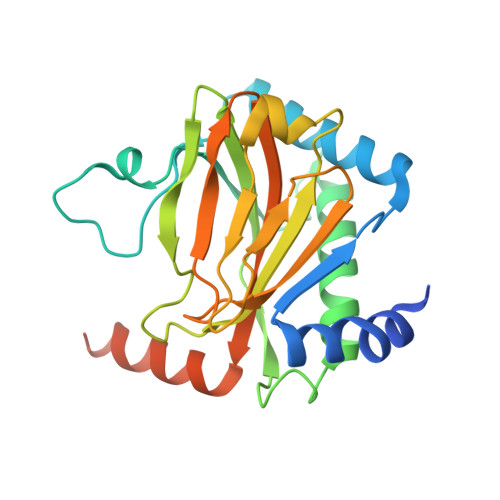
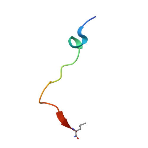
    Pacak-Zhuang syndrome is caused by mutations in the EPAS1 gene, which encodes for one of the three hypoxia-inducible factor alpha (HIFα) paralogs HIF2α and is associated with defined but varied phenotypic presentations including neuroendocrine tumors and polycythemia. However, the mechanisms underlying the complex genotype-phenotype correlations remain incompletely understood. Here, we devised a quantitative method for determining the dissociation constant (K d ) of the HIF2α peptides containing disease-associated mutations and the catalytic domain of prolyl-hydroxylase (PHD2) using microscale thermophoresis (MST) and showed that neuroendocrine-associated Class 1 HIF2α mutants have distinctly higher K d than the exclusively polycythemia-associated Class 2 HIF2α mutants. Based on the co-crystal structure of PHD2/HIF2α peptide complex at 1.8 Å resolution, we showed that the Class 1 mutated residues are localized to the critical interface between HIF2α and PHD2, adjacent to the PHD2 active catalytic site, while Class 2 mutated residues are localized to the more flexible region of HIF2α that makes less contact with PHD2. Concordantly, Class 1 mutations were found to significantly increase HIF2α-mediated transcriptional activation in cellulo compared to Class 2 counterparts. These results reveal a structural mechanism in which the strength of the interaction between HIF2α and PHD2 is at the root of the general genotype-phenotype correlations observed in Pacak-Zhuang syndrome.


  • Organizational Affiliation
    • Department of Laboratory Medicine & Pathobiology, Faculty of Medicine, University of Toronto, 1 King's College Circle, Toronto, ON, M5S 1A8, Canada.

Macromolecule Content 

  • Total Structure Weight: 30.42 kDa 
  • Atom Count: 2,014 
  • Modeled Residue Count: 240 
  • Deposited Residue Count: 271 
  • Unique protein chains: 2

Macromolecules

Find similar proteins by:|  3D Structure
Entity ID: 1
MoleculeChains  Sequence LengthOrganismDetailsImage
Egl nine homolog 1A [auth B]250Homo sapiensMutation(s): 0 
Gene Names: EGLN1C1orf12PNAS-118PNAS-137
EC: 1.14.11.29
UniProt & NIH Common Fund Data Resources
Find proteins for Q9GZT9 (Homo sapiens)
Explore Q9GZT9 
Go to UniProtKB:  Q9GZT9
PHAROS:  Q9GZT9
GTEx:  ENSG00000135766 
Entity Groups
Sequence Clusters30% Identity50% Identity70% Identity90% Identity95% Identity100% Identity
UniProt GroupQ9GZT9
Sequence Annotations
Expand
Reference Sequence
Find similar proteins by:  Sequence   |   3D Structure  
Entity ID: 2
MoleculeChains  Sequence LengthOrganismDetailsImage
Endothelial PAS domain-containing protein 1B [auth A]21Homo sapiensMutation(s): 1 
UniProt & NIH Common Fund Data Resources
Find proteins for Q99814 (Homo sapiens)
Explore Q99814 
Go to UniProtKB:  Q99814
PHAROS:  Q99814
GTEx:  ENSG00000116016 
Entity Groups
Sequence Clusters30% Identity50% Identity70% Identity90% Identity95% Identity100% Identity
UniProt GroupQ99814
Sequence Annotations
Expand
Reference Sequence

Small Molecules

Binding Affinity Annotations 
IDSourceBinding Affinity
OGA BindingDB:  7UJV Ki: 8000 (nM) from 1 assay(s)
Kd: min: 2000, max: 3400 (nM) from 5 assay(s)
IC50: min: 5600, max: 2.60e+4 (nM) from 6 assay(s)

Experimental Data & Validation

Experimental Data

  • Method: X-RAY DIFFRACTION
  • Resolution: 1.80 Å
  • R-Value Free:  0.213 (Depositor), 0.213 (DCC) 
  • R-Value Work:  0.173 (Depositor), 0.173 (DCC) 
Space Group: P 21 21 2
Unit Cell:
Length ( Å )Angle ( ˚ )
a = 129.078α = 90
b = 37.597β = 90
c = 42.148γ = 90
Software Package:
Software NamePurpose
REFMACrefinement
DIALSdata reduction
DIALSdata scaling
PHASERphasing

Structure Validation

View Full Validation Report


Ligand Structure Quality Assessment 


Entry History 

& Funding Information

Deposition Data


Funding OrganizationLocationGrant Number
Canadian Institutes of Health Research (CIHR)CanadaPJT-159773
Canada Research ChairsCanadaCRC-2017-00140
Canada Foundation for InnovationCanada--

Revision History  (Full details and data files)

  • Version 1.0: 2023-04-05
    Type: Initial release
  • Version 1.1: 2023-10-25
    Changes: Data collection, Refinement description
  • Version 1.2: 2024-03-13
    Changes: Database references
  • Version 1.3: 2024-11-13
    Changes: Structure summary