7UGW | pdb_00007ugw

M. tuberculosis DNA gyrase cleavage core bound to DNA and evybactin


Experimental Data Snapshot

  • Method: X-RAY DIFFRACTION
  • Resolution: 3.00 Å
  • R-Value Free: 
    0.276 (Depositor), 0.277 (DCC) 
  • R-Value Work: 
    0.218 (Depositor), 0.219 (DCC) 
  • R-Value Observed: 
    0.220 (Depositor) 

Starting Model: experimental
View more details

wwPDB Validation 3D Report Full Report

Validation slider image for 7UGW

This is version 2.0 of the entry. See complete history

Literature

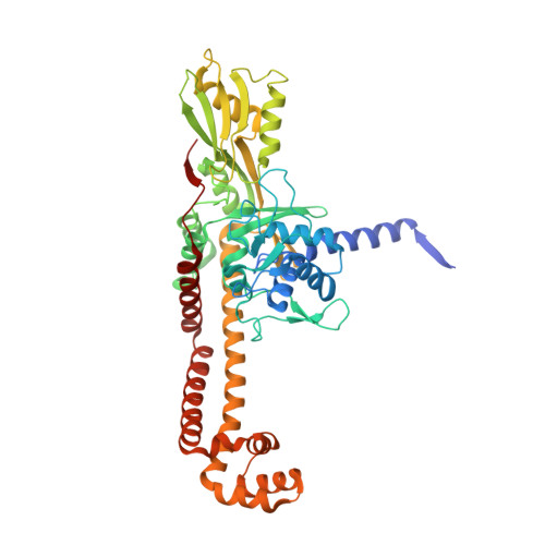
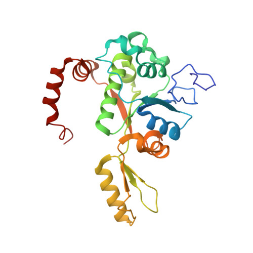
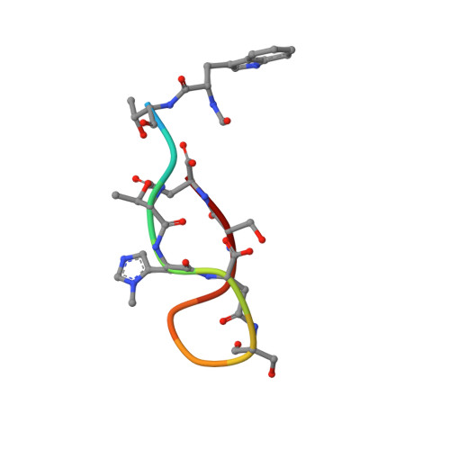
Evybactin is a DNA gyrase inhibitor that selectively kills Mycobacterium tuberculosis.

Imai, Y.Hauk, G.Quigley, J.Liang, L.Son, S.Ghiglieri, M.Gates, M.F.Morrissette, M.Shahsavari, N.Niles, S.Baldisseri, D.Honrao, C.Ma, X.Guo, J.J.Berger, J.M.Lewis, K.

(2022) Nat Chem Biol 18: 1236-1244

  • DOI: https://doi.org/10.1038/s41589-022-01102-7
  • Primary Citation Related Structures: 
    7UGW

  • PubMed Abstract: 

    The antimicrobial resistance crisis requires the introduction of novel antibiotics. The use of conventional broad-spectrum compounds selects for resistance in off-target pathogens and harms the microbiome. This is especially true for Mycobacterium tuberculosis, where treatment requires a 6-month course of antibiotics. Here we show that a novel antimicrobial from Photorhabdus noenieputensis, which we named evybactin, is a potent and selective antibiotic acting against M. tuberculosis. Evybactin targets DNA gyrase and binds to a site overlapping with synthetic thiophene poisons. Given the conserved nature of DNA gyrase, the observed selectivity against M. tuberculosis is puzzling. We found that evybactin is smuggled into the cell by a promiscuous transporter of hydrophilic compounds, BacA. Evybactin is the first, but likely not the only, antimicrobial compound found to employ this unusual mechanism of selectivity.


  • Organizational Affiliation
    • Antimicrobial Discovery Center, Department of Biology, Northeastern University, Boston, MA, USA.

Macromolecule Content 

  • Total Structure Weight: 183.78 kDa 
  • Atom Count: 12,374 
  • Modeled Residue Count: 1,510 
  • Deposited Residue Count: 1,560 
  • Unique protein chains: 3
  • Unique nucleic acid chains: 1

Macromolecules


Find similar proteins by:|  3D Structure
Entity ID: 1
MoleculeChains  Sequence LengthOrganismDetailsImage
DNA gyrase subunit A
A, C
500Mycobacterium tuberculosis H37RvMutation(s): 1 
Gene Names: gyrARv0006MTCY10H4.04
EC: 5.6.2.2
UniProt
Find proteins for P9WG47 (Mycobacterium tuberculosis (strain ATCC 25618 / H37Rv))
Explore P9WG47 
Go to UniProtKB:  P9WG47
Entity Groups
Sequence Clusters30% Identity50% Identity70% Identity90% Identity95% Identity100% Identity
UniProt GroupP9WG47
Sequence Annotations
Expand
Reference Sequence
Find similar proteins by:|  3D Structure
Entity ID: 2
MoleculeChains  Sequence LengthOrganismDetailsImage
DNA gyrase subunit B
B, D
251Mycobacterium tuberculosis H37RvMutation(s): 0 
Gene Names: gyrBRv0005MTCY10H4.03
EC: 5.6.2.2
UniProt
Find proteins for P9WG45 (Mycobacterium tuberculosis (strain ATCC 25618 / H37Rv))
Explore P9WG45 
Go to UniProtKB:  P9WG45
Entity Groups
Sequence Clusters30% Identity50% Identity70% Identity90% Identity95% Identity100% Identity
UniProt GroupP9WG45
Sequence Annotations
Expand
Reference Sequence
Find similar proteins by:  Sequence   |   3D Structure  
Entity ID: 4
MoleculeChains  Sequence LengthOrganismDetailsImage
evybactinF [auth E]12Photorhabdus noenieputensisMutation(s): 0 
Find similar nucleic acids by:  (by identity cutoff) 
Entity ID: 3
MoleculeChains LengthOrganismImage
DNA (46-MER)E [auth V]46synthetic construct
Sequence Annotations
Expand
Reference Sequence

Small Molecules

Modified Residues  1 Unique
IDChains TypeFormula2D DiagramParent
A1AZ2
Query on A1AZ2
F [auth E]D-PEPTIDE LINKINGC7 H11 N3 O2HIS
DAR
Query on DAR
F [auth E]D-PEPTIDE LINKINGC6 H15 N4 O2

--

DSN
Query on DSN
F [auth E]D-PEPTIDE LINKINGC3 H7 N O3

--

Biologically Interesting Molecules (External Reference) 

1 Unique

Experimental Data & Validation

Experimental Data

  • Method: X-RAY DIFFRACTION
  • Resolution: 3.00 Å
  • R-Value Free:  0.276 (Depositor), 0.277 (DCC) 
  • R-Value Work:  0.218 (Depositor), 0.219 (DCC) 
  • R-Value Observed: 0.220 (Depositor) 
Space Group: P 21 21 21
Unit Cell:
Length ( Å )Angle ( ˚ )
a = 83.075α = 90
b = 105.088β = 90
c = 250.183γ = 90
Software Package:
Software NamePurpose
PHENIXrefinement
Cootmodel building
XDSdata reduction
Aimlessdata scaling
PHASERphasing

Structure Validation

View Full Validation Report


Entry History 

& Funding Information

Deposition Data


Funding OrganizationLocationGrant Number
National Institutes of Health/National Institute Of Allergy and Infectious Diseases (NIH/NIAID)United StatesP01AI118687
National Institutes of Health/National Cancer Institute (NIH/NCI)United StatesR01CA077373
National Institutes of Health/National Cancer Institute (NIH/NCI)United StatesR35263778

Revision History  (Full details and data files)

  • Version 1.0: 2022-08-17
    Type: Initial release
  • Version 1.1: 2022-09-07
    Changes: Database references
  • Version 1.2: 2022-11-09
    Changes: Database references
  • Version 1.3: 2023-10-18
    Changes: Data collection, Refinement description
  • Version 2.0: 2024-10-02
    Type: Coordinate replacement
    Reason: Chirality error
    Changes: Advisory, Atomic model, Author supporting evidence, Data collection, Derived calculations, Non-polymer description, Other, Polymer sequence, Refinement description, Structure summary