Discovery of Nanomolar DCAF1 Small Molecule Ligands.
Li, A.S.M., Kimani, S., Wilson, B., Noureldin, M., Gonzalez-Alvarez, H., Mamai, A., Hoffer, L., Guilinger, J.P., Zhang, Y., von Rechenberg, M., Disch, J.S., Mulhern, C.J., Slakman, B.L., Cuozzo, J.W., Dong, A., Poda, G., Mohammed, M., Saraon, P., Mittal, M., Modh, P., Rathod, V., Patel, B., Ackloo, S., Santhakumar, V., Szewczyk, M.M., Barsyte-Lovejoy, D., Arrowsmith, C.H., Marcellus, R., Guie, M.A., Keefe, A.D., Brown, P.J., Halabelian, L., Al-Awar, R., Vedadi, M.(2023) J Med Chem 66: 5041-5060
- PubMed: 36948210 Search on PubMedSearch on PubMed Central
- DOI: https://doi.org/10.1021/acs.jmedchem.2c02132
- Primary Citation Related Structures:
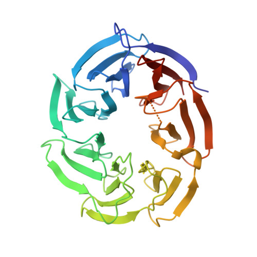
7UFV - PubMed Abstract:
DCAF1 is a substrate receptor of two distinct E3 ligases (CRL4 DCAF1 and EDVP), plays a critical physiological role in protein degradation, and is considered a drug target for various cancers. Antagonists of DCAF1 could be used toward the development of therapeutics for cancers and viral treatments. We used the WDR domain of DCAF1 to screen a 114-billion-compound DNA encoded library (DEL) and identified candidate compounds using similarity search and machine learning. This led to the discovery of a compound (Z1391232269) with an SPR K D of 11 μM. Structure-guided hit optimization led to the discovery of OICR-8268 ( 26e ) with an SPR K D of 38 nM and cellular target engagement with EC 50 of 10 μM as measured by cellular thermal shift assay (CETSA). OICR-8268 is an excellent tool compound to enable the development of next-generation DCAF1 ligands toward cancer therapeutics, further investigation of DCAF1 functions in cells, and the development of DCAF1-based PROTACs.
- Structural Genomics Consortium, University of Toronto, Toronto, Ontario M5G 1L7, Canada.
Organizational Affiliation: