Development and Optimization of Bifunctional Fusion Proteins to Locally Modulate Complement Activation in Diseased Tissue.
Fahnoe, K.C., Liu, F., Morgan, J.G., Ryan, S.T., Storek, M., Stark, E.G., Taylor, F.R., Holers, V.M., Thurman, J.M., Wawersik, S., Kalled, S.L., Violette, S.M.(2022) Front Immunol 13: 869725-869725
- PubMed: 35784298 Search on PubMedSearch on PubMed Central
- DOI: https://doi.org/10.3389/fimmu.2022.869725
- Primary Citation Related Structures:
7UE9 - PubMed Abstract:
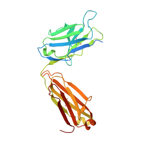
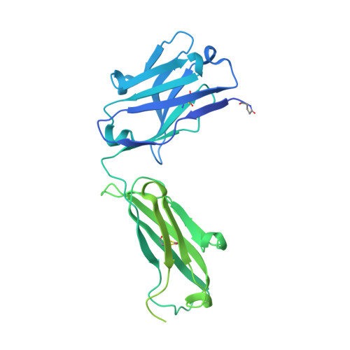
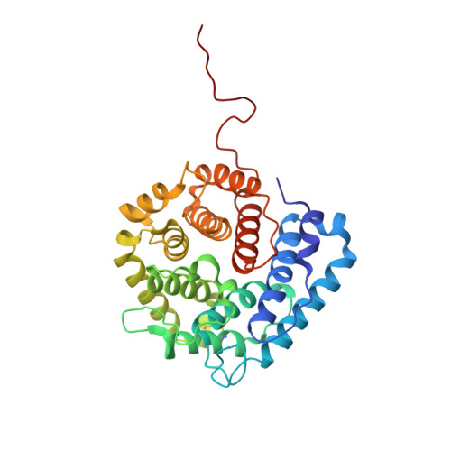
Sustained complement activation is an underlying pathologic driver in many inflammatory and autoimmune diseases. Currently approved anti-complement therapies are directed at the systemic blockade of complement. Consequently, these therapies provide widespread inhibition of complement pathway activity, beyond the site of ongoing activation and the intended pharmacodynamic (PD) effects. Given the essential role for complement in both innate and adaptive immunity, there is a need for therapies that inhibit complement in diseased tissue while limiting systemic blockade. One potential approach focuses on the development of novel fusion proteins that enable tissue-targeted delivery of complement negative regulatory proteins. These therapies are expected to provide increased potency and prolonged tissue PD, decreased dosing frequency, and the potential for improved safety profiles. We created a library of bifunctional fusion proteins that direct a fragment of the complement negative regulator, complement receptor type 1 (CR1) to sites of tissue injury. Tissue targeting is accomplished through the binding of the fusion protein to complement C3 fragments that contain a surface-exposed C3d domain and which are covalently deposited on tissues where complement is being activated. To that end, we generated a fusion protein that contains an anti-C3d monoclonal antibody recombinantly linked to the first 10 consensus repeats of CR1 (CR1 1-10 ) with the intention of delivering high local concentrations of this complement negative regulatory domain to tissue-bound complement C3 fragments iC3b, C3dg and C3d. Biochemical and in vitro characterization identified several fusion proteins that inhibit complement while maintaining the C3d domain binding properties of the parent monoclonal antibody. Preclinical in vivo studies further demonstrate that anti-C3d fusion proteins effectively distribute to injured tissue and reduce C3 fragment deposition for periods beyond 14 days. The in vitro and in vivo profiles support the further evaluation of C3d mAb-CR1 1-10 as a novel approach to restore proper complement activation in diseased tissue in the absence of continuous systemic complement blockade.
- Preclinical Research Q32 Bio Inc., Waltham, MA, United States.
Organizational Affiliation: