7UDU

cryo-EM structure of the ADP state wild type myosin-15-F-actin complex (symmetry expansion and re-centering)

  • Classification: MOTOR PROTEIN
  • Organism(s): Gallus gallus, Mus musculus
  • Expression System: Insect cell expression vector pTIE1
  • Mutation(s): No 

  • Deposited: 2022-03-20 Released: 2022-08-03 
  • Deposition Author(s): Gong, R., Reynolds, M.J., Alushin, G.M.
  • Funding Organization(s): National Institutes of Health/National Institute of Dental and Craniofacial Research (NIH/NIDCR), National Institutes of Health/National Institute of General Medical Sciences (NIH/NIGMS), National Institutes of Health/National Institute on Deafness and Other Communication Disorders (NIH/NIDCD)

Experimental Data Snapshot

  • Method: ELECTRON MICROSCOPY
  • Resolution: 4.15 Å
  • Aggregation State: FILAMENT 
  • Reconstruction Method: SINGLE PARTICLE 

wwPDB Validation   3D Report Full Report


This is version 1.0 of the entry. See complete history


Literature

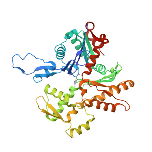
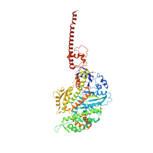
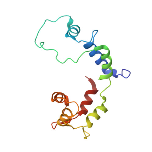
Structural basis for tunable control of actin dynamics by myosin-15 in mechanosensory stereocilia.

Gong, R.Jiang, F.Moreland, Z.G.Reynolds, M.J.de Los Reyes, S.E.Gurel, P.Shams, A.Heidings, J.B.Bowl, M.R.Bird, J.E.Alushin, G.M.

(2022) Sci Adv 8: eabl4733-eabl4733

  • DOI: https://doi.org/10.1126/sciadv.abl4733
  • Primary Citation of Related Structures:  
    7R8V, 7R91, 7RB8, 7RB9, 7UDT, 7UDU

  • PubMed Abstract: 

    The motor protein myosin-15 is necessary for the development and maintenance of mechanosensory stereocilia, and mutations in myosin-15 cause hereditary deafness. In addition to transporting actin regulatory machinery to stereocilia tips, myosin-15 directly nucleates actin filament ("F-actin") assembly, which is disrupted by a progressive hearing loss mutation (p.D1647G, " jordan "). Here, we present cryo-electron microscopy structures of myosin-15 bound to F-actin, providing a framework for interpreting the impacts of deafness mutations on motor activity and actin nucleation. Rigor myosin-15 evokes conformational changes in F-actin yet maintains flexibility in actin's D-loop, which mediates inter-subunit contacts, while the jordan mutant locks the D-loop in a single conformation. Adenosine diphosphate-bound myosin-15 also locks the D-loop, which correspondingly blunts actin-polymerization stimulation. We propose myosin-15 enhances polymerization by bridging actin protomers, regulating nucleation efficiency by modulating actin's structural plasticity in a myosin nucleotide state-dependent manner. This tunable regulation of actin polymerization could be harnessed to precisely control stereocilium height.


  • Organizational Affiliation

    Laboratory of Structural Biophysics and Mechanobiology, The Rockefeller University, New York, NY, USA.


Macromolecules
Find similar proteins by:  (by identity cutoff)  |  3D Structure
Entity ID: 1
MoleculeChains Sequence LengthOrganismDetailsImage
Actin, alpha skeletal muscle
A, B, C
373Gallus gallusMutation(s): 0 
UniProt
Find proteins for P68139 (Gallus gallus)
Explore P68139 
Go to UniProtKB:  P68139
Entity Groups  
Sequence Clusters30% Identity50% Identity70% Identity90% Identity95% Identity100% Identity
UniProt GroupP68139
Sequence Annotations
Expand
  • Reference Sequence
Find similar proteins by:  (by identity cutoff)  |  3D Structure
Entity ID: 2
MoleculeChains Sequence LengthOrganismDetailsImage
Unconventional myosin-XV719Mus musculusMutation(s): 0 
Gene Names: Myo15aMyo15
UniProt & NIH Common Fund Data Resources
Find proteins for Q9QZZ4 (Mus musculus)
Explore Q9QZZ4 
Go to UniProtKB:  Q9QZZ4
IMPC:  MGI:1261811
Entity Groups  
Sequence Clusters30% Identity50% Identity70% Identity90% Identity95% Identity100% Identity
UniProt GroupQ9QZZ4
Sequence Annotations
Expand
  • Reference Sequence
Find similar proteins by:  (by identity cutoff)  |  3D Structure
Entity ID: 3
MoleculeChains Sequence LengthOrganismDetailsImage
regulatory light chain146Gallus gallusMutation(s): 0 
UniProt
Find proteins for P24032 (Gallus gallus)
Explore P24032 
Go to UniProtKB:  P24032
Entity Groups  
Sequence Clusters30% Identity50% Identity70% Identity90% Identity95% Identity100% Identity
UniProt GroupP24032
Sequence Annotations
Expand
  • Reference Sequence
Small Molecules
Modified Residues  1 Unique
IDChains TypeFormula2D DiagramParent
HIC
Query on HIC
A, B, C
L-PEPTIDE LINKINGC7 H11 N3 O2HIS
Experimental Data & Validation

Experimental Data

  • Method: ELECTRON MICROSCOPY
  • Resolution: 4.15 Å
  • Aggregation State: FILAMENT 
  • Reconstruction Method: SINGLE PARTICLE 
EM Software:
TaskSoftware PackageVersion
MODEL REFINEMENTPHENIX
RECONSTRUCTIONRELION3.0

Structure Validation

View Full Validation Report


Entry History & Funding Information

Deposition Data


Funding OrganizationLocationGrant Number
National Institutes of Health/National Institute of Dental and Craniofacial Research (NIH/NIDCR)United StatesDP5OD017885
National Institutes of Health/National Institute of General Medical Sciences (NIH/NIGMS)United StatesR01GM141044
National Institutes of Health/National Institute on Deafness and Other Communication Disorders (NIH/NIDCD)United StatesR01DC018827

Revision History  (Full details and data files)

  • Version 1.0: 2022-08-03
    Type: Initial release