7UBU

Crystal structure of ZMET2 in complex with hemimethylated CAG DNA and a histone H3Kc9me2 peptide


Experimental Data Snapshot

  • Method: X-RAY DIFFRACTION
  • Resolution: 2.39 Å
  • R-Value Free: 0.256 
  • R-Value Work: 0.211 
  • R-Value Observed: 0.213 

wwPDB Validation   3D Report Full Report


Ligand Structure Quality Assessment 


This is version 1.3 of the entry. See complete history


Literature

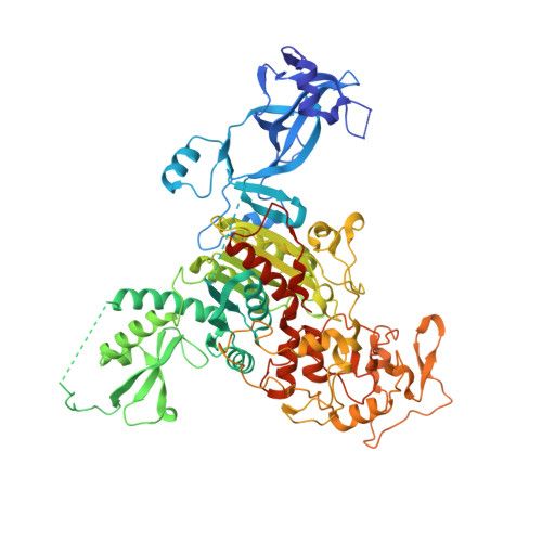
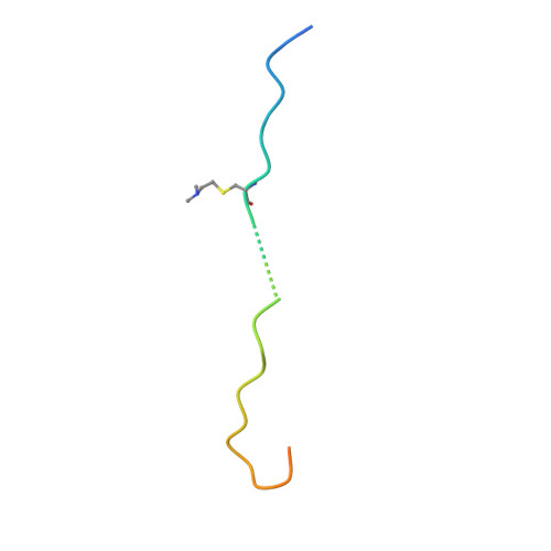
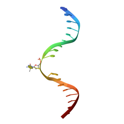
Mechanistic basis for maintenance of CHG DNA methylation in plants.

Fang, J.Jiang, J.Leichter, S.M.Liu, J.Biswal, M.Khudaverdyan, N.Zhong, X.Song, J.

(2022) Nat Commun 13: 3877-3877

  • DOI: https://doi.org/10.1038/s41467-022-31627-3
  • Primary Citation of Related Structures:  
    7UBU

  • PubMed Abstract: 

    DNA methylation is an evolutionarily conserved epigenetic mechanism essential for transposon silencing and heterochromatin assembly. In plants, DNA methylation widely occurs in the CG, CHG, and CHH (H = A, C, or T) contexts, with the maintenance of CHG methylation mediated by CMT3 chromomethylase. However, how CMT3 interacts with the chromatin environment for faithful maintenance of CHG methylation is unclear. Here we report structure-function characterization of the H3K9me2-directed maintenance of CHG methylation by CMT3 and its Zea mays ortholog ZMET2. Base-specific interactions and DNA deformation coordinately underpin the substrate specificity of CMT3 and ZMET2, while a bivalent readout of H3K9me2 and H3K18 allosterically stimulates substrate binding. Disruption of the interaction with DNA or H3K9me2/H3K18 led to loss of CMT3/ZMET2 activity in vitro and impairment of genome-wide CHG methylation in vivo. Together, our study uncovers how the intricate interplay of CMT3, repressive histone marks, and DNA sequence mediates heterochromatic CHG methylation.


  • Organizational Affiliation

    Department of Biochemistry, University of California, Riverside, CA, 92521, USA.


Macromolecules

Find similar proteins by:  (by identity cutoff)  |  3D Structure
Entity ID: 1
MoleculeChains Sequence LengthOrganismDetailsImage
DNA (cytosine-5)-methyltransferase 1783Zea maysMutation(s): 0 
Gene Names: MET2A
EC: 2.1.1.37
UniProt
Find proteins for Q9AXT8 (Zea mays)
Explore Q9AXT8 
Go to UniProtKB:  Q9AXT8
Entity Groups  
Sequence Clusters30% Identity50% Identity70% Identity90% Identity95% Identity100% Identity
UniProt GroupQ9AXT8
Sequence Annotations
Expand
  • Reference Sequence
Find similar proteins by:  (by identity cutoff)  |  3D Structure
Entity ID: 2
MoleculeChains Sequence LengthOrganismDetailsImage
Histone H3.2B [auth P],
E [auth Q]
33Zea maysMutation(s): 0 
Gene Names: H3C2H3C3H3C4
UniProt
Find proteins for P69246 (Zea mays)
Explore P69246 
Go to UniProtKB:  P69246
Entity Groups  
Sequence Clusters30% Identity50% Identity70% Identity90% Identity95% Identity100% Identity
UniProt GroupP69246
Sequence Annotations
Expand
  • Reference Sequence

Find similar nucleic acids by:  Sequence   |   3D Structure  

Entity ID: 3
MoleculeChains LengthOrganismImage
5MC SSDNAC [auth B]18Zea mays
Sequence Annotations
Expand
  • Reference Sequence

Find similar nucleic acids by:  Sequence   |   3D Structure  

Entity ID: 4
MoleculeChains LengthOrganismImage
C49 SSDNAD [auth C]18Zea mays
Sequence Annotations
Expand
  • Reference Sequence
Small Molecules
Ligands 1 Unique
IDChains Name / Formula / InChI Key2D Diagram3D Interactions
SAH (Subject of Investigation/LOI)
Query on SAH

Download Ideal Coordinates CCD File 
F [auth A]S-ADENOSYL-L-HOMOCYSTEINE
C14 H20 N6 O5 S
ZJUKTBDSGOFHSH-WFMPWKQPSA-N
Modified Residues  1 Unique
IDChains TypeFormula2D DiagramParent
M2L
Query on M2L
B [auth P],
E [auth Q]
L-PEPTIDE LINKINGC7 H16 N2 O2 SLYS
Experimental Data & Validation

Experimental Data

  • Method: X-RAY DIFFRACTION
  • Resolution: 2.39 Å
  • R-Value Free: 0.256 
  • R-Value Work: 0.211 
  • R-Value Observed: 0.213 
  • Space Group: P 21 21 21
Unit Cell:
Length ( Å )Angle ( ˚ )
a = 64.574α = 90
b = 121.392β = 90
c = 160.446γ = 90
Software Package:
Software NamePurpose
HKL-3000data scaling
PHENIXrefinement
PDB_EXTRACTdata extraction
Cootmodel building
PHASERphasing
HKL-3000data reduction

Structure Validation

View Full Validation Report


Ligand Structure Quality Assessment 


Entry History & Funding Information

Deposition Data


Funding OrganizationLocationGrant Number
National Institutes of Health/National Institute of General Medical Sciences (NIH/NIGMS)United States1R35GM119721

Revision History  (Full details and data files)

  • Version 1.0: 2022-06-08
    Type: Initial release
  • Version 1.1: 2022-07-20
    Changes: Database references
  • Version 1.2: 2023-10-18
    Changes: Data collection, Refinement description
  • Version 1.3: 2023-11-15
    Changes: Data collection