Crystal structures of pertussis toxin with NAD + and analogs provide structural insights into the mechanism of its cytosolic ADP-ribosylation activity.
Sakari, M., Tran, M.T., Rossjohn, J., Pulliainen, A.T., Beddoe, T., Littler, D.R.(2022) J Biological Chem 298: 101892-101892
- PubMed: 35378130 Search on PubMedSearch on PubMed Central
- DOI: https://doi.org/10.1016/j.jbc.2022.101892
- Primary Citation Related Structures:
7SKI, 7SKK, 7SKY, 7SNE, 7U6Z - PubMed Abstract:
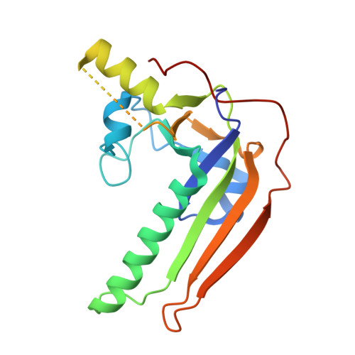
Bordetella pertussis is the causative agent of whooping cough, a highly contagious respiratory disease. Pertussis toxin (PT), a major virulence factor secreted by B. pertussis, is an AB5-type protein complex topologically related to cholera toxin. The PT protein complex is internalized by host cells and follows a retrograde trafficking route to the endoplasmic reticulum, where it subsequently dissociates. The released enzymatic S1 subunit is then translocated from the endoplasmic reticulum into the cytosol and subsequently ADP-ribosylates the inhibitory alpha-subunits (Gαi) of heterotrimeric G proteins, thus promoting dysregulation of G protein-coupled receptor signaling. However, the mechanistic details of the ADP-ribosylation activity of PT are not well understood. Here, we describe crystal structures of the S1 subunit in complex with nicotinamide adenine dinucleotide (NAD+), with NAD+ hydrolysis products ADP-ribose and nicotinamide, with NAD+ analog PJ34, and with a novel NAD+ analog formed upon S1 subunit crystallization with 3-amino benzamide and NAD+, which we name benzamide amino adenine dinucleotide. These crystal structures provide unprecedented insights into pre- and post-NAD+ hydrolysis steps of the ADP-ribosyltransferase activity of PT. We propose that these data may aid in rational drug design approaches and further development of PT-specific small-molecule inhibitors.
- Institute of Biomedicine, Research Unit for Infection and Immunity, University of Turku, Turku, Finland.
Organizational Affiliation: